. 2010 Jun 11;285(24):18452-63.
doi: 10.1074/jbc.M109.077925. Epub 2010 Mar 5.

Aldehyde dehydrogenase 7A1 (ALDH7A1) is a novel enzyme involved in cellular defense against hyperosmotic stress

Affiliations

Aldehyde dehydrogenase 7A1 (ALDH7A1) is a novel enzyme involved in cellular defense against hyperosmotic stress

Chad Brocker et al. J Biol Chem. .

Abstract

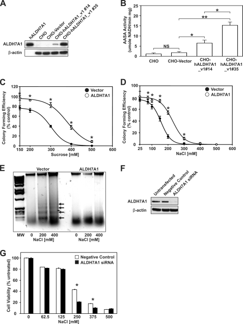
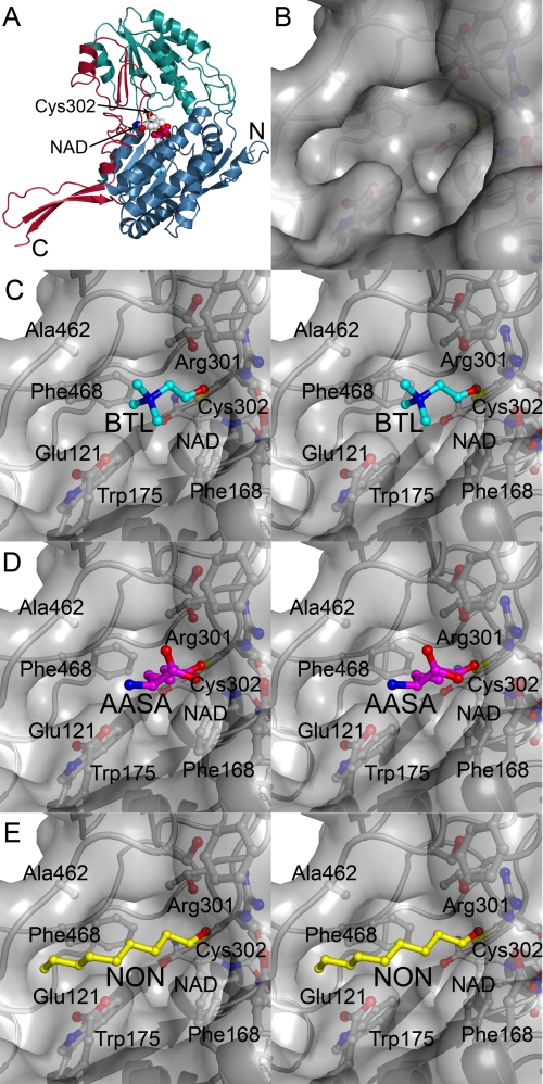
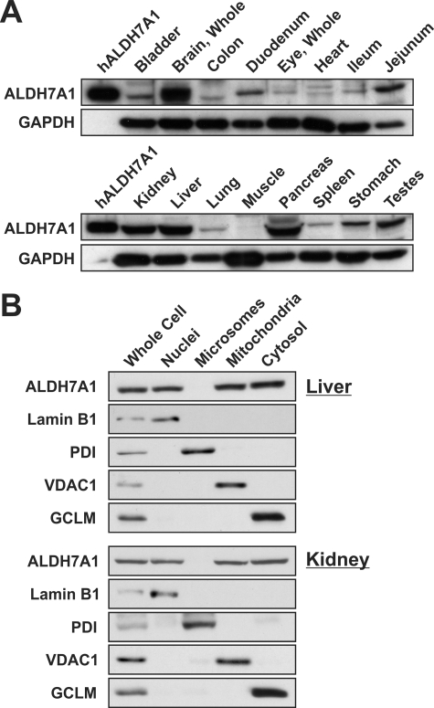
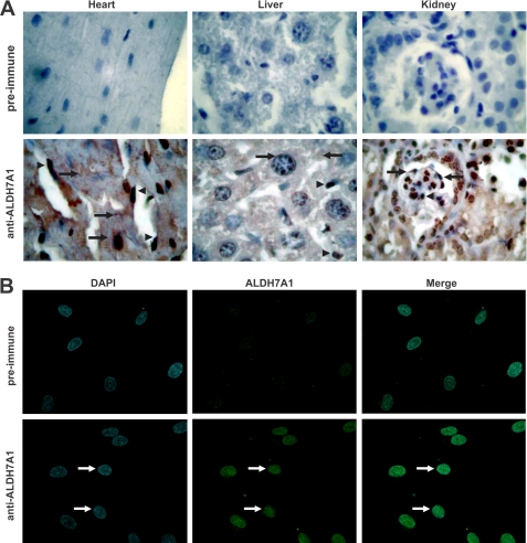
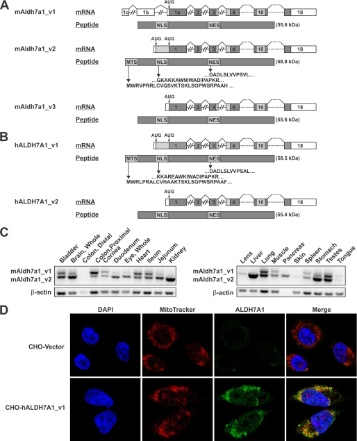
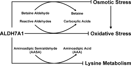
Mammalian ALDH7A1 is homologous to plant ALDH7B1, an enzyme that protects against various forms of stress, such as salinity, dehydration, and osmotic stress. It is known that mutations in the human ALDH7A1 gene cause pyridoxine-dependent and folic acid-responsive seizures. Herein, we show for the first time that human ALDH7A1 protects against hyperosmotic stress by generating osmolytes and metabolizing toxic aldehydes. Human ALDH7A1 expression in Chinese hamster ovary cells attenuated osmotic stress-induced apoptosis caused by increased extracellular concentrations of sucrose or sodium chloride. Purified recombinant ALDH7A1 efficiently metabolized a number of aldehyde substrates, including the osmolyte precursor, betaine aldehyde, lipid peroxidation-derived aldehydes, and the intermediate lysine degradation product, alpha-aminoadipic semialdehyde. The crystal structure for ALDH7A1 supports the enzyme's substrate specificities. Tissue distribution studies in mice showed the highest expression of ALDH7A1 protein in liver, kidney, and brain, followed by pancreas and testes. ALDH7A1 protein was found in the cytosol, nucleus, and mitochondria, making it unique among the aldehyde dehydrogenase enzymes. Analysis of human and mouse cDNA sequences revealed mitochondrial and cytosolic transcripts that are differentially expressed in a tissue-specific manner in mice. In conclusion, ALDH7A1 is a novel aldehyde dehydrogenase expressed in multiple subcellular compartments that protects against hyperosmotic stress by generating osmolytes and metabolizing toxic aldehydes.


Figures

FIGURE 1.
FIGURE 1.
Expression of ALDH7A1 in CHO cells protects against hyperosmotic stress. A, cell lysates (30 μg) from untransfected CHO cells and CHO cell lines stably transfected with ΔpCEP4Δ vector (CHO-Vector) or ΔpCEP4Δ-hALDH7A1_v1 (CHO-hALDH7A1_v1#13 and CHO-hALDH7A1_v1#35) were assayed for ALDH7A1 protein expression by Western blot analysis, using specific antibodies (as described under “Experimental Procedures”; top). The blot was reprobed for β-actin as loading correction. Purified recombinant human ALDH7A1 protein (50 ng) was used as positive control. B, cell lysates were assayed for AASA activity (as described under “Experimental Procedures”). C and D, CHO-Vector (filled circles) and CHO-hALDH7A1_v1 clone 35 (open circles) cells were treated with sucrose (C) or NaCl (D) for 4 h at 37 °C, and colonies were stained with crystal violet after 1 week. Mean colony numbers obtained with CHO-Vector and CHO-hALDH7A1_v1 clone 35 cells were normalized as a percentage of untreated control. E, CHO-Vector (Vector) and CHO-hALDH7A1_v1 clone 35 (ALDH7A1) cells were treated with 200 or 400 mm NaCl for 4 h at 37 °C. Agarose gel electrophoresis was performed on DNA isolated from untreated and treated cells. Only in CHO-Vector cells did NaCl promote the characteristic 200-bp DNA fragment laddering associated with apoptotic cell death (arrows). F, Western analysis of ALDH7A1 knockdown in HK-2 cell lysates (20 μg). Cells were transfected with 20 nm negative control or ALDH7A1-specific siRNA. Untransfected cells were used as an additional negative control. Blots were reprobed for β-actin. G, decreased cell viability in HK-2 cells lacking ALDH7A1. siRNA-transfected cells were treated with increasing concentrations of NaCl, and cell viability was analyzed via an MTT assay. Values represent mean ± S.E. (error bars) for triplicates in three separate experiments (NS, not significant (p > 0.05); *, p < 0.05; **, p < 0.01).
FIGURE 2.
FIGURE 2.
Human ALDH7A1 crystal structure and docked substrates. A, α-carbon trace of human ALDH7A1 subunit A with the N-terminal cofactor binding domain (blue), the C-terminal catalytic domain (green), and the oligomerization domain (red). The NAD molecule is shown in spheres and the catalytic Cys302 is shown in a ball-and stick representation at the interface of the two larger domains. B, surface representation of active-site pocket. C–E are colored according to atom type with nitrogen blue, oxygen red, sulfur yellow, and protein carbon gray; stereoview close-up of betaine aldehyde (BTL; cyan carbons) (C), AASA (magenta carbons) (D), and nonanal (NON; yellow carbons) (E) docked into the active site. The orientation is the same as in A.
FIGURE 3.
FIGURE 3.
Tissue and subcellular distribution of mouse ALDH7A1. Individual organs were isolated from adult C57BL/6J/129/SvJ mice and prepared for Western blot analysis of ALDH7A1 expression (as described under “Experimental Procedures”). A, tissue distribution of ALDH7A1. One hundred ng of purified human ALDH7A1 protein was loaded into lane 1 (left lane, hALDH7A1), and 30 μg of protein-soluble extract obtained from a variety of tissues was loaded into each additional lane. The same membranes were reprobed for glyceraldehyde-3-phosphate dehydrogenase (bottom panels) for loading correction. B, ALDH7A1 expression in subcellular fractions from liver and kidney was determined via Western blot analysis (as described under “Experimental Procedures”). Ten μg of whole cell or subcellular fraction lysate was loaded into each lane. Blots were probed with lamin B1 (Nuclei), protein-disulfide isomerase (Microsomes), VDAC1 (Mitochondria), and GCLM (Cytosol) to assess fraction purity.
FIGURE 4.
FIGURE 4.
ALDH7A1 immunochemical staining in mouse tissues and primary HUVEC. A, heart, liver, and kidney were isolated from C57BL/6J/129/SvJ adult mice. Five-μm sections (obtained and processed as described under “Experimental Procedures”) were incubated with preimmune serum (top) or human ALDH7A1 antibody (bottom), followed by streptavidin-conjugated goat anti-rabbit IgG secondary antibody. Representative staining patterns are shown at ×400 magnification. Arrowheads, intense nuclear staining in endothelial cells of all tissues. Additionally, strong ALDH7A1 staining was detected in the nuclei and cytosol of cardiomyocytes and hepatocytes as well as podocyte nuclei, in heart, liver, and kidney tissues sections, respectively (arrows). B, HUVEC grown in culture and probed with human ALDH7A1 antibody followed by fluorescein isothiocyanate-conjugated secondary antibody (green). Nuclei were stained with DAPI (blue). ALDH7A1 staining is predominantly nuclear (arrows). Co-localization was visualized by overlaying DAPI and ALDH7A1 images (Merge).
FIGURE 5.
FIGURE 5.
Identification of mitochondrial ALDH7A1 transcript. A, diagram illustrating alternative splicing event at the 5′-end of the mouse Aldh7a1 gene, which gives rise to two splice variants, mAldh7a1_v1 and mAldh7a1_v2. Alternative splicing results in the removal of upstream AUG, which subsequently prevents translation of a mitochondrial localization signal. Another transcript, mAldh7a1_v3, lacks upstream exons present in mAldh7a1_v1 but utilizes the same start codon. B, the human ALDH7A1 gene contains two potential translation initiation sites. hALDH7A1_v1 and hALDH7A1_v2 have been sequenced and utilize different AUG start codons, resulting in either retention or removal of a mitochondrial targeting signal. Upstream AUG in both mAldh7a1_v2 and hALDH7A1_v1 are predicted to be non-optimal translation start sites, indicating potential use of a “leaky scanning” mechanism for translation initiation. To date, no homolog for mAldh7a1_v1 has been identified in humans. Both A and B also include predicted nuclear localization and nuclear export signals. Introns are not drawn to scale (MTS, mitochondrial targeting signal; NLS, nuclear localization signal; NES, nuclear export signal). C, RT-PCR of C57BL6 mouse tissues using splice variant-specific ALDH7A1 primers. Non-mitochondrial transcript is indicated by the 937 bp upper band. Mitochondrial transcript is identified by the 779 bp lower band. β-Actin primers were used as positive and loading controls. D, immunocytochemical staining of stable CHO cells transfected with mammalian expression vector comprising human ALDH7A1 containing the mitochondrial signal sequence (CHO-hALDH7A1_v1) or empty vector (CHO-Vector). Fixed cells were probed with polyclonal human ALDH7A1 antibody, followed by fluorescein isothiocyanate-conjugated secondary antibody (green). Mitochondria labeled with Invitrogen's MitoTracker® Orange CM-H2 TMRos probe (red). Nuclei were stained with DAPI (blue). The merged image denotes strong ALDH7A1 mitochondrial localization.
FIGURE 6.
FIGURE 6.
Proposed role of ALDH7A1 in the cell. ALDH7A1 protects cells and tissues from osmotic stress through the synthesis of the protective osmolyte betaine from betaine aldehyde. Furthermore, ALDH7A1 can directly metabolize a wide range of reactive aldehydes produced during lipid peroxidation, which potentiate oxidative stress within the cell. ALDH7A1 also plays a key role in lysine metabolism through the conversion of AASA to aminoadipic acid (AAA).

Similar articles

Cited by

References

    1. Marchitti S. A., Brocker C., Stagos D., Vasiliou V. (2008) Expert Opin. Drug Metab. Toxicol. 4, 697–720 - PMC - PubMed
    1. Lee P., Kuhl W., Gelbart T., Kamimura T., West C., Beutler E. (1994) Genomics 21, 371–378 - PubMed
    1. Malik H. S., Henikoff S. (2003) Nat. Struct. Biol. 10, 882–891 - PubMed
    1. Guerrero F. D., Jones J. T., Mullet J. E. (1990) Plant Mol. Biol. 15, 11–26 - PubMed
    1. Stroeher V. L., Boothe J. G., Good A. G. (1995) Plant Mol. Biol. 27, 541–551 - PubMed

Publication types

Associated data