. 2010 May 7;285(19):14521-33.
doi: 10.1074/jbc.M110.112409. Epub 2010 Mar 5.

Mutations of the opsin gene (Y102H and I307N) lead to light-induced degeneration of photoreceptors and constitutive activation of phototransduction in mice

Affiliations

Mutations of the opsin gene (Y102H and I307N) lead to light-induced degeneration of photoreceptors and constitutive activation of phototransduction in mice

Ewa Budzynski et al. J Biol Chem. .

Abstract

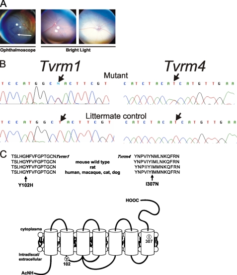
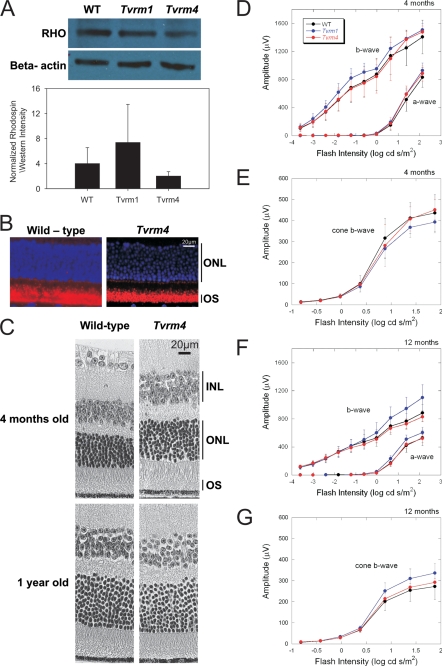
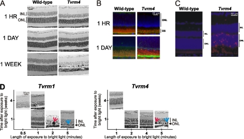
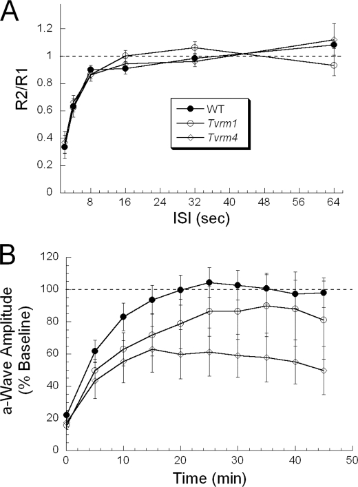
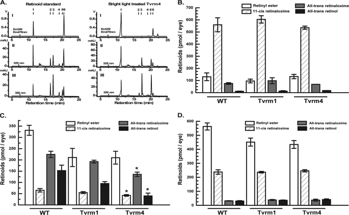
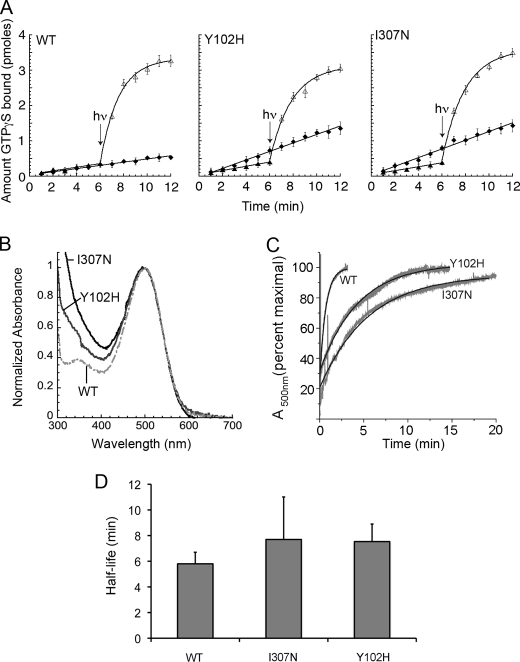
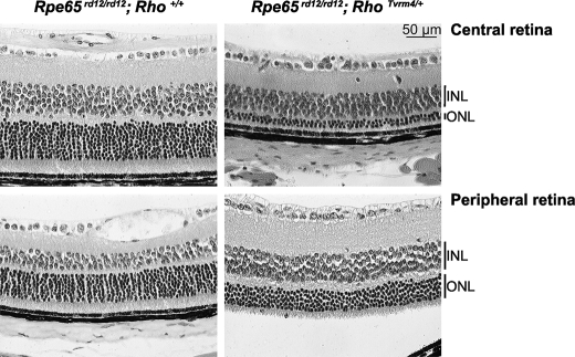
Mutations in the Rhodopsin (Rho) gene can lead to autosomal dominant retinitis pigmentosa (RP) in humans. Transgenic mouse models with mutations in Rho have been developed to study the disease. However, it is difficult to know the source of the photoreceptor (PR) degeneration in these transgenic models because overexpression of wild type (WT) Rho alone can lead to PR degeneration. Here, we report two chemically mutagenized mouse models carrying point mutations in Rho (Tvrm1 with an Y102H mutation and Tvrm4 with an I307N mutation). Both mutants express normal levels of rhodopsin that localize to the PR outer segments and do not exhibit PR degeneration when raised in ambient mouse room lighting; however, severe PR degeneration is observed after short exposures to bright light. Both mutations also cause a delay in recovery following bleaching. This defect might be due to a slower rate of chromophore binding by the mutant opsins compared with the WT form, and an increased rate of transducin activation by the unbound mutant opsins, which leads to a constitutive activation of the phototransduction cascade as revealed by in vitro biochemical assays. The mutant-free opsins produced by the respective mutant Rho genes appear to be more toxic to PRs, as Tvrm1 and Tvrm4 mutants lacking the 11-cis chromophore degenerate faster than mice expressing WT opsin that also lack the chromophore. Because of their phenotypic similarity to humans with B1 Rho mutations, these mutants will be important tools in examining mechanisms underlying Rho-induced RP and for testing therapeutic strategies.


Figures

FIGURE 1.
FIGURE 1.
Light-induced degeneration of photoreceptors in Tvrm4 and Tvrm1 mutants are caused by missense mutations in the Rhodopsin gene (Rho). A, fundus photographs of Tvrm4 mutant mice and controls. After light exposure from indirect ophthalmoscopy (left), the central retina becomes hyperpigmented, indicating photoreceptor degeneration, shown by a white arrow. Within 24 h after exposure to bright light (center and right panels) the mutant retina becomes hyper-reflective (right), whereas the WT retina does not (center). B, schematic of the Rho sequences depicting the Tvrm4 and Tvrm1 mutations. Single base pair substitutions (arrow) were found and predicted to lead to a change of amino acid 307, isoleucine to asparagine (I307N) 102 in Tvrm4, and tyrosine to histidine (Y102H) in Tvrm1, n = 3 animals. C, the amino acid residues, Ile-307 and Tyr-102, are conserved among species. Protein sequences are from different species containing Ile-307 and Tyr-102 amino acid residues. The sites of mutations are indicated in the rhodopsin crystal structure (PDB coordinates 1U19) using the molecular modeling program UCSF Chimera.
FIGURE 2.
FIGURE 2.
Photoreceptor degeneration does not occur in Tvrm4 and Tvrm1 Rho mutant mice raised in ambient light. A, Western analysis of rhodopsin levels in the retinas of WT and RhoTvrm4/+ mice. β-Actin is used as a loading control. Protein levels were quantified using ImageGauge. The rhodposin levels were normalized to the level of β-actin and are plotted as mean ± S.E. There were no significant differences between groups, one-way ANOVA, n = 3 mice. B, immunohistochemical analysis of rhodopsin in retinas of RhoTvrm4/+ mutant and WT animals raised in ambient light. Rhodopsin localization in the outer segments of Tvrm4 mutants (left) is not different from WT (right). Blue, 4′,6-diamidino-2-phenylindole staining; red, anti-rhodopsin staining, n = 3 mice. C, light microscopy of central retinas from aged animals. Photoreceptor nuclei of 4-month and 1-year-old RhoTvrm4/+ (right) animals raised in standard ambient mouse room light are preserved, comparable with wild-type littermates (left), n = 3 mice per group. D-G, intensity-response functions for ERGs obtained from WT mice and RhoTvrm1/+ and RhoTvrm4/+ mutants. ERGs were recorded at 4 (D and E) and 12 months (F and G) under dark-adapted (D and F) and light-adapted (E and G) conditions. Data points indicate the average ± S.E. for at least 5 mice. In comparison to WT littermates, there is no decrement in ERG amplitude in either RhoTvrm1/+ or RhoTvrm4/+ mice. INL, inner nuclear layer; ONL, outer nuclear layer; OS, outer segments of photoreceptors.
FIGURE 3.
FIGURE 3.
Photoreceptor degeneration in Tvrm1 and Tvrm4 mutants is rapidly induced by exposure to bright light. A, light microscopy and immunohistochemical staining of rhodopsin in the central retina from Tvrm4 mutant mice with 1- or 5-min exposures to bright light. Histological changes in the retinas of RhoTvrm4/+ mutants are observed within 1 h (top) following exposure to bright light. At 24 h (middle) following the exposure to bright light, the outer and inner segments of the RhoTvrm4/+ photoreceptors are disorganized. At 1 week (bottom) following exposure, the outer nuclear layer is reduced to two rows of cell bodies. B, rhodopsin (red), ezrin (yellow), and 4′,6-diamidino-2-phenylindole (blue) staining of the retinas from WT and RhoTvrm4/+ animals 1 or 24 h after exposure to bright light. Photoreceptor outer segments are engulfed within the RPE cells of mutant eyes at 1 h after exposure to bright light, n = 2 animals. C, photoreceptors of Rho mutants are apoptotic 24 h following exposure to bright light. TUNEL staining of the retina was 24 h following bright light exposure, n = 3 animals. D, the Tvrm1 mutation makes the retina more vulnerable to light damage than the Tvrm4 mutation. Light microscopy of the central retina at different times following different durations of exposure to bright light. Red arrows indicate chromatin aggregates and blue arrows indicate macrophages, n = 3 mice for all groups except for RhoTvrm1/+, where n = 6.
FIGURE 4.
FIGURE 4.
Recovery from a high intensity level bleach is impaired in the mutants but activation and deactivation of phototransduction are normal. A, paired-flash analysis of phototransduction deactivation. In each trial, responses obtained to the probe flash (R2) are expressed relative to the response obtained to the conditioning flash (R1) and are plotted as a function of the duration of the ISI separating the conditioning and probe flashes. Data points indicate the mean ± S.E. for at least 5 mice. Recovery kinetics of the a-wave in RhoTvrm1/+ and RhoTvrm4/+ mice are comparable with those of WT littermates. B, bleaching recovery. Response recovery of a-wave amplitude following exposure to a high intensity bleaching light. For each individual mouse, responses were normalized to the dark-adapted pre-bleach baseline. Data points indicate mean ± S.E. for 19 WT, 9 RhoTvrm1/+, and 9 RhoTvrm4/+ mice. Compared with WT mice, recovery of the a-wave from bleaching light is slower and incomplete in RhoTvrm1/+ and RhoTvrm4/+ mice.
FIGURE 5.
FIGURE 5.
HPLC analysis of visual cycle retinoids. A, typical HPLC chromatograms; 1, retinyl esters; 2, syn-11-cis-retinaloxime; 3, syn-all-trans-retinaloxime; 4, anti-11-cis-retinaloxime; 5, anti-all-trans-retinaloxime; 6, all-trans-retinol. Panel I, fluorescence detection with 325 nm excitation and 475 nm emission; II, detection of retinyl ester and retinol at 325 nm by photodiode array detector; III, detection of oximes at 350 nm by photodiode array. B, retinoid levels in dark adapted WT, Tvrm1, and Tvrm4 mice. Total amounts extracted, as described under “Materials and Methods,” are plotted in units of pmol/eye. C, retinoid levels immediately after exposure to bright light. Amounts extracted from eyes immediately after exposure to bright light are shown. D, recovery of retinoids 1 h after return to darkness. After treatment with bright light, animals were returned to darkness for 1 h prior to collection of eyes and extraction of retinoids. Data are mean ± S.E. p < 0.05 One-way ANOVA using the F-distribution for samples from all three genotypes followed by Dunnett's test, n = 4 samples of 3 eyes each.
FIGURE 6.
FIGURE 6.
In vitro biochemistry of mutant and WT rhodopsin. A, transducin activation by receptors in the presence and absence of chromophore is not impaired in the mutants. Closed diamonds, time course for the reaction catalyzed by the apoprotein opsin. Closed circles, time course for the reaction catalyzed by rhodopsin in the dark (all slopes shown are indistinguishable from those observed with transducin alone under these conditions). Open triangles, time course for the reaction catalyzed by rhodopsin after exposure to light (hν). The data are plotted as mean ± S.D. B, normalized dark absorbance spectra show maximum absorbance of mutants similar to that of WT rhodopsin. C, the mutant opsins bind chromophore more slowly than does WT opsin. Kinetics of retinal binding to Y102H (N2C, Y102H, D282C) and I307N (N2C, D282C, I307N) opsins are slowed relative to ET (N2C, D282C) opsin. Opsins were purified, and change in absorbance at 500 nm was recorded continuously after addition of a 5-fold molar excess of 11-cis-retinal. The absorbance values were normalized by the final maximum changes observed for each experiment. Representative raw data are depicted in gray, and the calculated best-fit curve for a single exponential in black. Mean lifetimes ± S.E. measured were as follows: 0.655 ± 0.014 min for WT, 4.32 ± 0.44 min for Y102H, and 5.94 ± 0.29 min for I307N (p < 0.0001, one-way ANOVA). D, mean half-lives of metarhodopsin II decay for WT (N2C, D282C), Y102H (N2C, Y102H, D282C), and I307N (N2C, D282C, I307N) rhodopsin. Rhodopsin, in the presence of a 3-fold molar excess of 11-cis-retinal, was exposed to white light passed through a 480-nm cut-on filter for 1 s, and the change of absorbance at 500 nm was monitored. Under these conditions, the rate-limiting step in regeneration is that of metarhodopsin II (3). The mean half-life of metarhodospin II decay of the mutants was not significantly different from the WT value, one-way ANOVA.
FIGURE 7.
FIGURE 7.
Mutant opsins lead to photoreceptor degeneration without exposure to bright light. Histological sections from central and peripheral retinas of 4-week-old Rpe65rd12/rd12,Rho+/+ and Rpe65rd12/rd12/RhoTvrm4/+ animals, n = 3 mice.

Similar articles

Cited by

References

    1. Rim J., Oprian D. D. (1995) Biochemistry 34, 11938–11945 - PubMed
    1. Li T., Franson W. K., Gordon J. W., Berson E. L., Dryja T. P. (1995) Proc. Natl. Acad. Sci. U.S.A. 92, 3551–3555 - PMC - PubMed
    1. Humphries M. M., Rancourt D., Farrar G. J., Kenna P., Hazel M., Bush R. A., Sieving P. A., Sheils D. M., McNally N., Creighton P., Erven A., Boros A., Gulya K., Capecchi M. R., Humphries P. (1997) Nat. Genet. 15, 216–219 - PubMed
    1. Lem J., Krasnoperova N. V., Calvert P. D., Kosaras B., Cameron D. A., Nicolò M., Makino C. L., Sidman R. L. (1999) Proc. Natl. Acad. Sci. U.S.A. 96, 736–741 - PMC - PubMed
    1. Toda K., Bush R. A., Humphries P., Sieving P. A. (1999) Vis. Neurosci. 16, 391–398 - PubMed

Publication types