. 2010 Mar;22(3):782-96.
doi: 10.1105/tpc.109.072173. Epub 2010 Mar 5.

bZIP28 and NF-Y transcription factors are activated by ER stress and assemble into a transcriptional complex to regulate stress response genes in Arabidopsis

Affiliations

bZIP28 and NF-Y transcription factors are activated by ER stress and assemble into a transcriptional complex to regulate stress response genes in Arabidopsis

Jian-Xiang Liu et al. Plant Cell. 2010 Mar.

Abstract

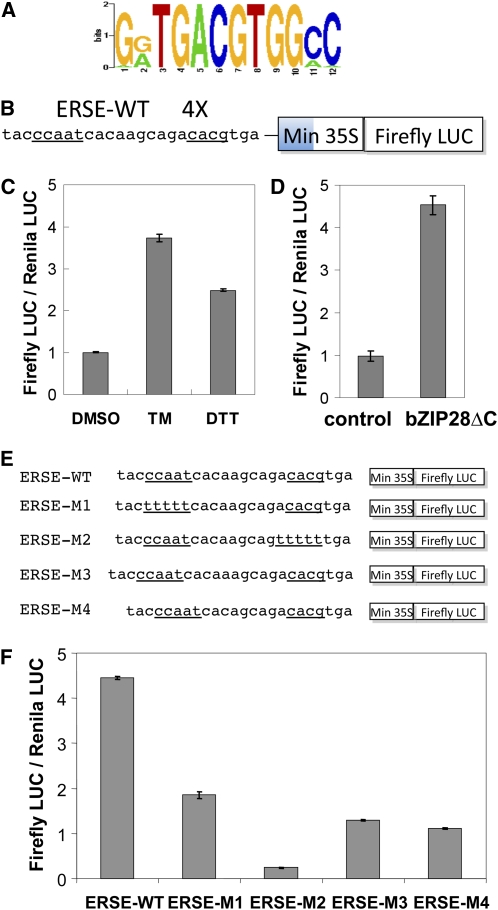
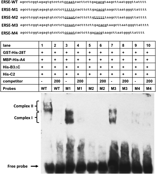
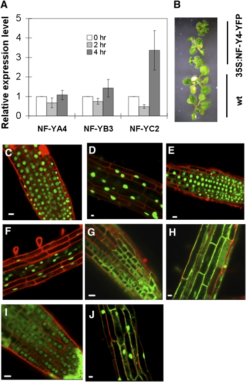
Stress agents known to elicit the unfolded protein response in Arabidopsis thaliana upregulate the expression of a constellation of genes dependent on the membrane-associated basic domain/leucine zipper (bZIP) transcription factor, bZIP28. Among the stress-activated genes, a consensus promoter sequence corresponding to the endoplasmic reticulum (ER) stress-responsive element I (ERSE-I), CCAAT-N10-CACG, was identified. Disruption of either the CCAAT or CACG subelement in ERSE-I resulted in reduction of the transcriptional response to ER stress. bZIP28 forms homo- and heterodimers with other bZIP TF family members (in subgroup D) and interacts with CCAAT box binding factors, heterotrimeric factors composed of NF-Y subunits. Arabidopsis encodes 36 NF-Y subunits, and it was found that subunits NF-YB3 and -YC2 interact with bZIP28 and NF-YA4, respectively, in a yeast three-hybrid system. A transcriptional complex containing bZIP28 and the above-mentioned three NF-Y subunits was assembled in vitro on DNA containing ERSE-I. bZIP28, on its own, binds to the CACG subelement in ERSE-I to form a smaller complex I, and in combination with the NF-Y subunits above, bZIP28 assembles into a larger transcriptional complex (complex II). bZIP28 was shown to interact with NF-Y subunits in vivo in bimolecular fluorescence complementation analyses and in coimmunoprecipitation assays. Treatment of seedlings with ER stress agents led to the upregulation of NF-YC2 and the relocation of NF-YB3 from the cytoplasm to the nucleus. Thus, in response to ER stress, bZIP28 is mobilized by proteolysis and recruits NF-Y subunits to form a transcriptional complex that upregulates the expression of ER stress-induced genes.


Figures

Figure 1.
Figure 1.
Genes Dependent on bZIP28 for UPR Upregulation. Wild-type and zip28-2 Arabidopsis seedlings were treated with 5 μg/mL TM or DMSO (mock treatment) for 2 h, and total RNA was extracted and analyzed by quantitative RT-PCR. Expression levels of BiP3 (At1g09080) (A), SHD (At4g24190) (B), CNX1 (At5g61790) (C), SDF2 (At2g25110) (D), SHP70 (At4g16660) (E), and PDIL1-1 (At1g21750) (F) were normalized to actin 2/8. The values in DMSO control were normalized to 1 (At3g18780/ At1g49240). Error bars represent se for three biological replicates. Differences between genotypes were statistically significant (P = 0.05) according to Tukey's range (honestly significant difference) test and two-way analysis of variance.
Figure 2.
Figure 2.
The ERSE-I Promoter Element Confers Responsiveness to ER Stress Agents in UPR-Induced Genes. (A) Consensus sequence for the common promoter element in bZIP28-dependent genes derived from PSCAN analysis. (B) A construct was developed containing (as shown) a 4X multimer of an ERSE-I promoter element from the BiP3 gene linked to a minimal CaMV 35S promoter and firefly luciferase (LUC). The underlined sequences are two core subelements in ERSE-I. (C) and (D) The construct was tested for induction in an Arabidopsis protoplast system treated with UPR stress agents, TM or DTT, or mock treated (DMSO) (C) or cotransfected with a constitutively active form of bZIP28ΔC or empty vector (control) (D). Test plasmids also encode a constitutively active Renilla LUC, and activity was expressed as a ratio of firefly LUC/Renilla LUC activity. According to a Student's t test, the treatment (C), bZIP28ΔC addition (D), and each ERSE-I mutation (F) were statistically significant (P = 0.05). (E) Mutated forms of the ERSE-I (ERSE-M1 through -M4) in which the CCAAT box or the bZIP binding site was multiply mutated by base substitution. (F) Activity of the mutated ERSE-I forms in Arabidopsis protoplasts was also expressed as a ratio of firefly LUC/Renilla LUC activity. Error bars represent se for three independent experiments.
Figure 3.
Figure 3.
Formation of Homo- and Heterodimers between bZIP TFs. The N-terminal segments of members of the D family of bZIP TFs (bZIP17, -28, -49, and -60) were tested as bait and prey in a yeast two-hybrid system. In the N-terminal segments, the transcription activation domain, transmembrane domain, and C-terminal regions were removed. (A) Growth of yeast on SD-Trp-Leu plates. (B) Growth of yeast on SD-Trp-Leu-His plates. Activation of HIS gene (shown by ability to grow on –His plates) was used as the indication of interaction.
Figure 4.
Figure 4.
Interaction of NF-Y Subunits with bZIP28. (A) The N-terminal segment of bZIP28 (bZIP28T) was fused to the Gal4 UAS binding domain (BD) and used as bait in a yeast three-hybrid system against a pool of NF-YB and -YC subunits. The transcription activation domain, transmembrane domain, and C-terminal region were removed in bZIP28T. Prey plasmids harboring NF-YB (prey B, with URA selection marker) and -YC (prey C, with TRP selection marker) subunit genes were pooled and challenged against bZIP28T as bait (with LEU selection marker) and tested for their ability to activate the HIS interaction marker. (B) The recovered plasmids from the screening were tested pairwise (one NF-YB and -YC subunit in each test) against bZIP28T as bait in the three-hybrid system. (C) NF-YA4 was used as bait in the three-hybrid system in a similar pairwise challenge with NF-Y subunits (-Trp-Leu-Ura were noninteraction selective conditions, and -Trp-Leu-Ura-His were interaction selective conditions). (D) NF-YA4 was used as bait in a two-hybrid system to test the interaction with prey NF-YB3 or NF-YC2. (E) NF-YC2 was used as bait in a two-hybrid system to investigate the potential for dimer formation. Activation of the HIS gene was used as the indication of interaction in (D) and (E).
Figure 5.
Figure 5.
Pull-Down Assay to Detect the in Vitro Assembly of a Transcriptional Complex on DNA Containing ERSE-I. DNA was incubated with an epitope-tagged, truncated form of bZIP28 (GST-H6-28T) and various epitope-tagged forms of NF-Y subunits (MBP-H6-NF-YA4, H6-NF-YB3ΔC, and H6-NF-YC2). The proteins were synthesized in E. coli, and the amounts of tagged NF-Y subunits (lanes contain 30% input material) added to the assembly reactions were estimated by protein immunoblot using anti-His antibody (lanes 8 to 10). Protein complexes containing GST-H6-28T were pulled down using glutathione agarose beads. Transcriptional complexes containing MBP-H6-NF-YA4 were detected with anti-MBP.
Figure 6.
Figure 6.
Electrophoretic Mobility Shift Assays to Detect the in Vitro Assembly of a Transcriptional Complex on DNA Containing ERSE-I. Biotinylated DNA containing ERSE-I was incubated with an epitope-tagged, truncated form of bZIP28 and various epitope-tagged forms of NF-Y subunits as described in Figure 5. To determine whether the assembly of the transcriptional complex was specific for DNA containing ERSE-I, 50- to 200-fold excess unlabeled competitor ERSE-I DNA was added to the reactions as indicated. The migration positions of complexes I and II are indicated. To demonstrate that the tagged proteins are involved in the assembly of complex II, the reactions were postincubated with anti-His antibody. Complex II* represents the resulting supershifted complex.
Figure 7.
Figure 7.
Effect of Modifications of the ERSE-I DNA on the Assembly of a bZIP28-Containing Transcriptional Complex. Various ERSE-I constructs (ERSE-M1 to -M2) were tested in electrophoretic mobility shift assays in which the CCAAT box or bZIP binding site was altered by multiple base substitutions or in which the length of the spacer was altered. Migration positions of complex I and II are indicated as described in Figure 6. The 200-fold excess unlabeled competitor DNA was added to the control reactions to determine whether binding is specific.
Figure 8.
Figure 8.
Detection the Interaction between bZIP28 and NF-Y Subunits in Vivo with BiFC and Pull-Down Assays. The BiFC assay involves the interaction of proteins fused to N-terminal (nYFP) and C-terminal (cYFP) fragments of YFP to restore fluorescence of the split fluorophore. (A) to (F) Individual NF-Y subunits were fused to nYFP, and the truncated form of bZIP28 (bZIP28T) was fused to cYFP. Each NF-Y subunit fusion protein was coexpressed with bZIP28T-cYFP in Arabidopsis roots. Roots were counterstained with 50 μg/mL propidium iodide (red). Bars = 20 μm. (A) to (C) Untreated (DMSO). (D) to (F) TM treated for 4 h. (A) and (D) nYFP-NF-YA4 plus bZIP28T-cYFP. (B) and (E) nYFP-NF-YB3 plus bZIP28T-cYFP. (C) and (F) nYFP-NF-YC2 plus bZIP28T-cYFP. (G) Pull-down assay with NF-YB3-YFP–overexpressing seedlings. Total proteins were extracted from nontreated and TM-treated (5 μg/mL) 2-week old seedlings and incubated with purified GST-His-bZIP28T (GST-His is a negative control). The complex was pulled down with glutathione beads and detected with anti-GFP antibody against YFP. Purified His-NF-YC2 was also added along with GST-His-bZIP28T in nontreated sample (lane 5). Two percent of the input material in the pull-down reactions was also loaded as a sample in lanes 6 and 7. The origin of doublet bands of NF-YB3-YFP is not known.
Figure 9.
Figure 9.
Induction of NF-Y Gene Expression and Subcellular Relocation of NF-Y Subunits. (A) Quantitative RT-PCR analysis of the expression of NF-Y subunits following TM treatment. (B) NF-YA4-YFP overexpression plants were smaller than wild-type plants at 2 weeks. (C) to (J) Merged confocal microscopy images of YFP-tagged NF-Y subunits in Arabidopsis root tips ([C], [E], [G], and [I]) or in elongated cells in the root ([D], [F], [H], and [J]). Samples were counterstained with propidium iodide (red). (C) and (D) NF-YA4-YFP, untreated (DMSO). (E) and (F) NF-YC2-YFP, untreated. (G) and (H) NF-YB3-YFP, untreated. (I) and (J) NF-YB3-YFP, TM treated for 4 h. Bars = 10 mm in (B) and 10 μm in (C) to (J). Error bars in (A) represent se of three biological replicates. The relative expression level at 0 h was normalized to 1. Only the treatment for NF-YC2 is statistically significant (P = 0.05) according to Student's t test.
Figure 10.
Figure 10.
Model for the Assembly of bZIP28 and NF-Y Containing Transcriptional Complexes and the Upregulation of UPR Genes. In response to stress, bZIP28p is proteolytically activated by Golgi-localized S1P (and probably also by S2P) to release bZIP28n, which relocates to the nucleus to form transcriptional complexes on target genes (BiP3 and NF-YC2) bearing ERSEs in their promoters. Although the steps in nuclear translocation of the NF-Y subunits have not been studied in detail in plants, we infer from the mammalian cell literature (Kahle et al., 2005) that NF-YC2 dimerization with NF-YB3 allows the latter to be translocated to the nucleus and for the pair (NF-YC2 and NF-YB3) to interact with NF-YA4, which likely enters independently. Transcriptional complexes are composed of NF-Y trimers, which bind to CCAAT box subelements of the ERSEs, and bZIP28n dimers, which bind to bZIP binding subelements.

References

    1. Bellorini M., Lee D.K., Dantonel J.C., Zemzoumi K., Roeder R.G., Tora L., Mantovani R. (1997). CCAAT binding NF-Y-TBP interactions: NF-YB and NF-YC require short domains adjacent to their histone fold motifs for association with TBP basic residues. Nucleic Acids Res. 25: 2174–2181 - PMC - PubMed
    1. Ben-Naim O., Eshed R., Parnis A., Teper-Bamnolker P., Shalit A., Coupland G., Samach A., Lifschitz E. (2006). The CCAAT binding factor can mediate interactions between CONSTANS-like proteins and DNA. Plant J. 46: 462–476 - PubMed
    1. Bernales S., Papa F.R., Walter P. (2006). Intracellular signaling by the unfolded protein response. Annu. Rev. Cell Dev. Biol. 22: 487–508 - PubMed
    1. Cai X., Ballif J., Endo S., Davis E., Liang M., Chen D., DeWald D., Kreps J., Zhu T., Wu Y. (2007). A putative CCAAT-binding transcription factor is a regulator of flowering timing in Arabidopsis. Plant Physiol. 145: 98–105 - PMC - PubMed
    1. Chen X., Shen J., Prywes R. (2002). The luminal domain of ATF6 senses endoplasmic reticulum (ER) stress and causes translocation of ATF6 from the ER to the Golgi. J. Biol. Chem. 277: 13045–13052 - PubMed

Publication types

MeSH terms

Substances