. 2010 Jun;298(6):L744-54.
doi: 10.1152/ajplung.00368.2009. Epub 2010 Mar 5.

Zinc modulates the innate immune response in vivo to polymicrobial sepsis through regulation of NF-kappaB

Affiliations

Zinc modulates the innate immune response in vivo to polymicrobial sepsis through regulation of NF-kappaB

Shengying Bao et al. Am J Physiol Lung Cell Mol Physiol. 2010 Jun.

Abstract

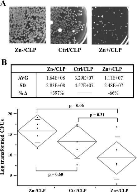
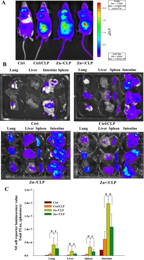
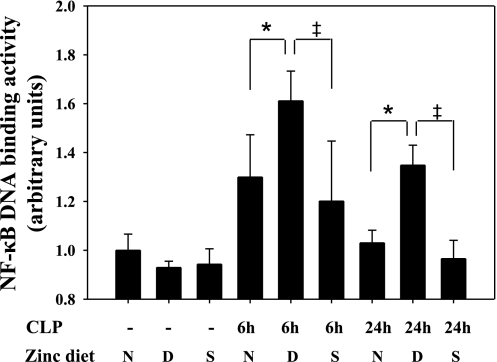
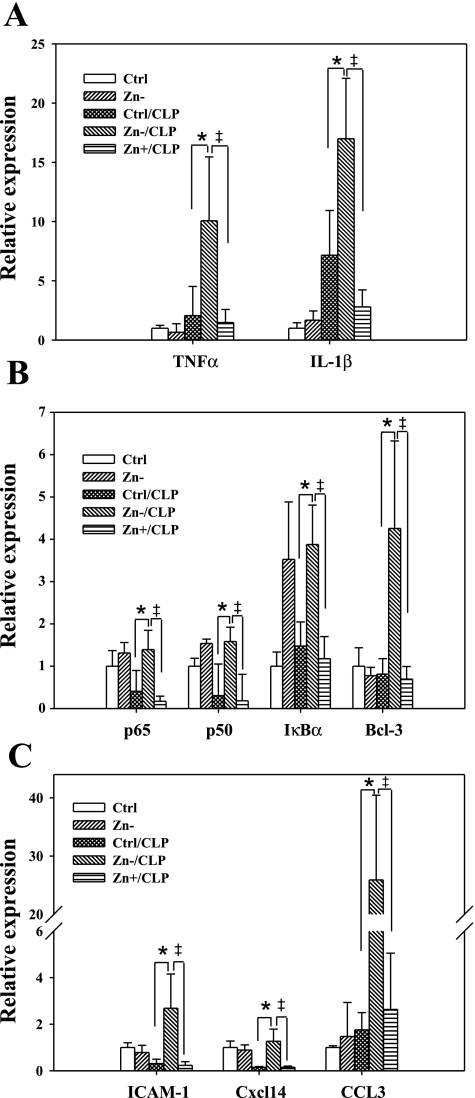
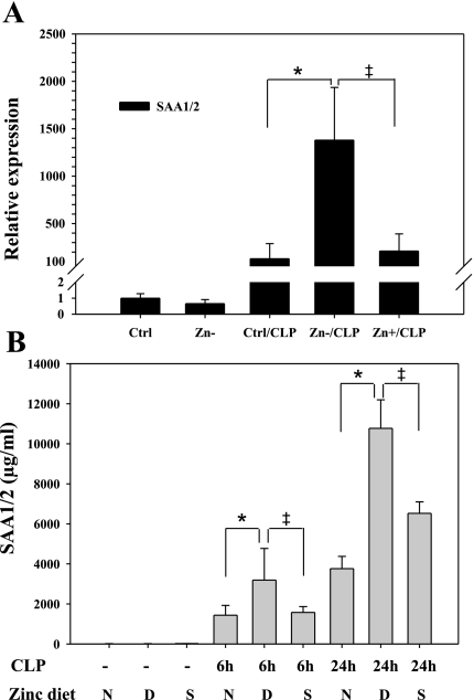
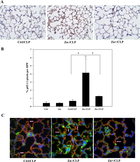
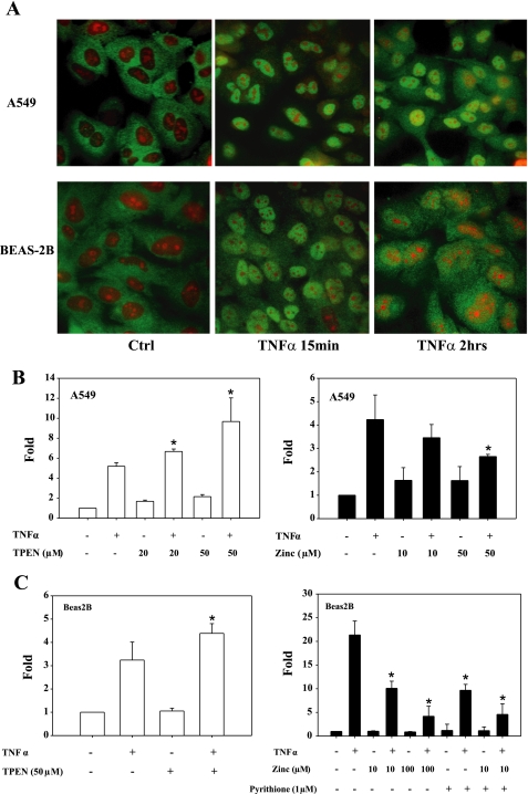
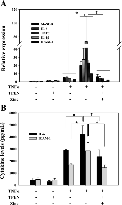
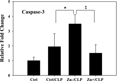
Zinc is an essential element that facilitates coordination of immune activation during the host response to infection. We recently reported that zinc deficiency increases systemic inflammation, vital organ damage, and mortality in a small animal model of sepsis. To investigate potential mechanisms that cause these phenomena, we used the same animal model and observed that zinc deficiency increases bacterial burden and enhances NF-kappaB activity in vital organs including the lung. We conducted further studies in the lung to determine the overall impact of zinc deficiency. At the molecular level, NF-kappaB p65 DNA-binding activity was enhanced by zinc deficiency in response to polymicrobial sepsis. Furthermore, expression of the NF-kappaB-targeted genes IL-1beta, TNFalpha, ICAM-1, and the acute phase response gene SAA1/2 were elevated by zinc deficiency. Unexpectedly, the amount of NF-kappaB p65 mRNA and protein was increased in the lung including alveolar epithelia of zinc-deficient mice. These events occurred with a significant and concomitant increase in caspase-3 activity within 24 h of sepsis onset in zinc-deficient mice relative to control group. Short-term zinc supplementation reversed these effects. Reconstitution of zinc deficiency in lung epithelial cultures resulted in similar findings in response to TNFalpha. Taken together, zinc deficiency systemically enhances the spread of infection and NF-kappaB activation in vivo in response to polymicrobial sepsis, leading to enhanced inflammation, lung injury, and, as reported previously, mortality. Zinc supplementation immediately before initiation of sepsis reversed these effects thereby supporting the plausibility of future studies that explore zinc supplementation strategies to prevent sepsis-mediated morbidity and mortality.


Figures

Fig. 1.
Fig. 1.
Zinc deficiency increases bacterial burden following cecal ligation and puncture (CLP). Whole blood was obtained 24 h following CLP treatment, plated on blood agar following serial dilutions, and then incubated overnight and enumerated the following day. A: representative pictures of plates from the zinc-deficient (Zn/CLP), zinc-sufficient (Ctrl/CLP), and zinc-supplemented (Zn+/CLP) treatment groups are shown. B, top: total bacterial counts were obtained from all animals in each treatment group. Average (AVG) and standard deviation (SD) are displayed in this table (n = 5 mice in each treatment group). %Δ Represents the % difference between the zinc-deficient (Zn) or zinc-supplemented (Zn+) compared with the baseline control zinc diet (Ctrl). B, bottom: bacterial counts were log transformed to obtain a normal distribution and then displayed graphically (long horizontal line in the diamond = group mean, and short horizontal lines in the diamond = SD). The differences between all groups were assessed by ANOVA (P = 0.07) followed by the Tukey honestly significant difference test to look for trends between each group, P values are shown. No bacteria were present in animals placed on normal or zinc-modified diets (data not shown). This figure is representative of 2 separate experiments. cfu, Colony-forming units.
Fig. 2.
Fig. 2.
Zinc deficiency increases NF-κB activation systemically following CLP. A: a representative whole body image obtained from 1 mouse within each treatment group is shown and includes the untreated control diet (Ctrl) group along with analysis of mice 8 h after CLP in conjunction with 3 different zinc diets, including the control diet (Ctrl/CLP), a zinc-deficient diet (Zn/CLP), or a zinc-deficient diet followed by acute zinc supplementation (Zn+/CLP) (n = 2 for untreated mice, n = 3 per dietary groups involving CLP treatment). The anesthetized mice were placed in a light-sealed chamber connected to the charge-coupled device (CCD) camera for image analysis. Luminescence emitted from each animal was integrated for 10 min starting 2 min after d-luciferin injection. B: vital organs including the liver, lung, spleen, and intestine were excised from each animal and immediately subjected to quantitative bioluminescent imaging. The composite images of each tissue from each animal are presented after color scale adjustment (the scale for A and B is identical). C: metric analysis of total photon flux of pooled data for each tissue from each treatment group (*P < 0.05, #not statistically significant).
Fig. 3.
Fig. 3.
Zinc deficiency increases NF-κB DNA-binding activity in vivo in the lung following CLP. DNA-binding activity of the transcriptional factor NF-κB p65 was quantified in mouse lung tissue obtained from adult male C57BL/6 mice placed on zinc-modified diets at 6 and 24 h following CLP. Values (means ± SD) were normalized according to cellular protein content. Data are representative of 3 separate experiments (n = 5 animals per treatment group: N, zinc-normal diet; D, zinc-deficient diet; S, short-term zinc supplementation diet after a zinc-deficient regimen). *Statistically significant difference between Zn/CLP and Ctrl/CLP, P < 0.05; ‡statistically significant difference between Zn+/CLP and Zn/CLP, P < 0.05.
Fig. 4.
Fig. 4.
Zinc deficiency modulates NF-κB-mediated related gene expression in the lung following sepsis. C57BL/6 black mice were subjected to modified diets for 3 wk and then subjected to CLP. Mouse lungs were lavaged and perfused with saline at 24 h following CLP. Total RNA was isolated, and the expression of NF-κB-related genes was determined by real-time PCR. The genes included: the cytokines IL-1β and TNFα (A); NF-κB family p65, p50, IκBα, and IκB family member B cell lymphoma 3 (Bcl-3) (B); and chemokines CCL3 and CXCL14 and the cell adhesion molecule ICAM-1 (C). n = 5 lungs per treatment group; *Zn/CLP vs. Ctrl/CLP, P < 0.05; ‡Zn+/CLP vs. Zn/CLP, P < 0.05.
Fig. 5.
Fig. 5.
Zinc deficiency enhances the acute phase response in response to sepsis. A: the mRNA levels of serum amyloid A 1/2 (SAA1/2) were measured by real-time PCR (n = 5 animals per treatment group). B: blood was obtained from the same animals at 6 or 24 h following CLP, and SAA levels were measured by ELISA. n = 5 per group; *Zn/CLP vs. Ctrl/CLP, P < 0.05; ‡Zn+/CLP vs. Zn/CLP, P < 0.05.
Fig. 6.
Fig. 6.
Increased expression of NF-κB p65 in the lung in response to zinc deficiency and CLP was found. Mouse lungs were inflated and fixed with formaldehyde immediately following lavage and perfusion. A: immunostaining was performed to detect p65 protein (magnification, ×200). Data are representative of a minimum of 5 mice per treatment group. B: 10 random images of each lung specimen were captured and analyzed. The number of brown pixels staining positive for total p65 protein was counted and compared between each treatment group (n = 5 per group; *Zn/CLP vs. Ctrl/CLP, P < 0.05; ‡Zn+/CLP vs. Zn/CLP, P < 0.05). C: immunofluorescent staining for p65 was also conducted in conjunction with confocal analysis to identify the cellular location of p65 (green) within lung parenchymal tissue. Rhodamine-labeled Ricinus communis agglutinin (RCA), a lectin-based stain for type I alveolar epithelial cell-specific RCA (red), as well as the nuclear stain 4′,6′-diamidino-2-phenylindole (DAPI; blue) were used to clarify the location, identity, and extent of p65-positive cells. Pink arrows designate p65-positive type I alveolar epithelia, whereas white arrows designate alveolar macrophages. HPF, high-powered field.
Fig. 7.
Fig. 7.
Zinc modulates NF-κB activity in human lung epithelia. A: TNFα (10 ng/ml) exposure lead to the rapid translocation of NF-κB into the nucleus of human lung epithelial cell lines A549 and BEAS-2B cells as determined by immunofluorescent detection of p65 protein (green). Propidium iodide was used to counterstain nuclei (red). B and C: zinc modulates NF-κB transactivation of a luciferase reporter gene (3×κB-luc) in A549 and BEAS-2B cells. Both cultures were transfected in triplicate with an NF-κB reporter plasmid (3×κB-luc). N,N,N′,N′-tetrakis(2-pyridylmethyl)ethylenediamine (TPEN; 20 or 50 μM × 1 h) was then added to deplete intracellular zinc. Cells were then exposed to TNFα (10 ng/ml) for 6 h. Zinc inhibition study was done in combination with pyrithione before addition of TNFα (10 ng/ml) for 1 h (*P < 0.05 compared with the group of TNFα treatment only).
Fig. 8.
Fig. 8.
Zinc deficiency enhances the expression of NF-κB-responsive genes in primary human lung epithelial cells. A: primary human lung epithelial cells (hLECs) were cultured onto collagen-coated, semipermeable membranes. TPEN (20 μM) or zinc (5 μM) were used to modify zinc status as previously described. Cells were then stimulated with TNFα (10 ng/ml) for 6 h to activate the NF-κB pathway. Total RNA was isolated, and the expression of NF-κB target genes was measured. B: in addition, culture supernatants were obtained and measured to determine IL-6 and ICAM-1 concentrations (total donor number: n = 3; *Zn/CLP vs. Ctrl/CLP, P < 0.05; ‡Zn+/CLP vs. Zn/CLP, P < 0.05). MnSOD, manganese superoxide dismutase.
Fig. 9.
Fig. 9.
Zinc deficiency increases caspase-3 activity in the lung in response to CLP. Identical lung specimens as described previously were processed and subjected to analysis for caspase-3 activity at 24 h after CLP in response to zinc deficiency as well as supplementation (n = 5 per group; *Zn/CLP vs. Ctrl/CLP, P < 0.05; ‡Zn+/CLP vs. Zn/CLP, P < 0.05).

References

    1. Abraham E, Matthay MA, Dinarello CA, Vincent JL, Cohen J, Opal SM, Glauser M, Parsons P, Fisher CJ, Jr, Repine JE. Consensus conference definitions for sepsis, septic shock, acute lung injury, and acute respiratory distress syndrome: time for a reevaluation. Crit Care Med 28: 232–235, 2000 - PubMed
    1. Abraham E, Singer M. Mechanisms of sepsis-induced organ dysfunction. Crit Care Med 35: 2408–2416, 2007 - PubMed
    1. Balibrea JL, Arias-Diaz J. Acute respiratory distress syndrome in the septic surgical patient. World J Surg 27: 1275–1284, 2003 - PubMed
    1. Bao S, Knoell DL. Zinc modulates airway epithelium susceptibility to death receptor-mediated apoptosis. Am J Physiol Lung Cell Mol Physiol 290: L433–L441, 2006 - PubMed
    1. Bohrer H, Qiu F, Zimmermann T, Zhang Y, Jllmer T, Mannel D, Bottiger BW, Stern DM, Waldherr R, Saeger HD, Ziegler R, Bierhaus A, Martin E, Nawroth PP. Role of NFkappaB in the mortality of sepsis. J Clin Invest 100: 972–985, 1997 - PMC - PubMed

Publication types

MeSH terms