. 2010 May;24(5):1007-23.
doi: 10.1210/me.2009-0472. Epub 2010 Mar 5.

Androgens induce nongenomic stimulation of colonic contractile activity through induction of calcium sensitization and phosphorylation of LC20 and CPI-17

Affiliations

Androgens induce nongenomic stimulation of colonic contractile activity through induction of calcium sensitization and phosphorylation of LC20 and CPI-17

María C González-Montelongo et al. Mol Endocrinol. 2010 May.

Abstract

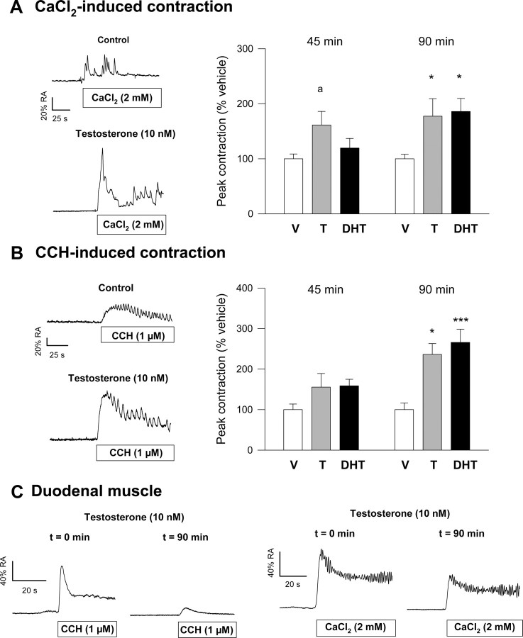
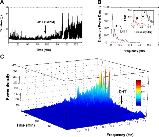
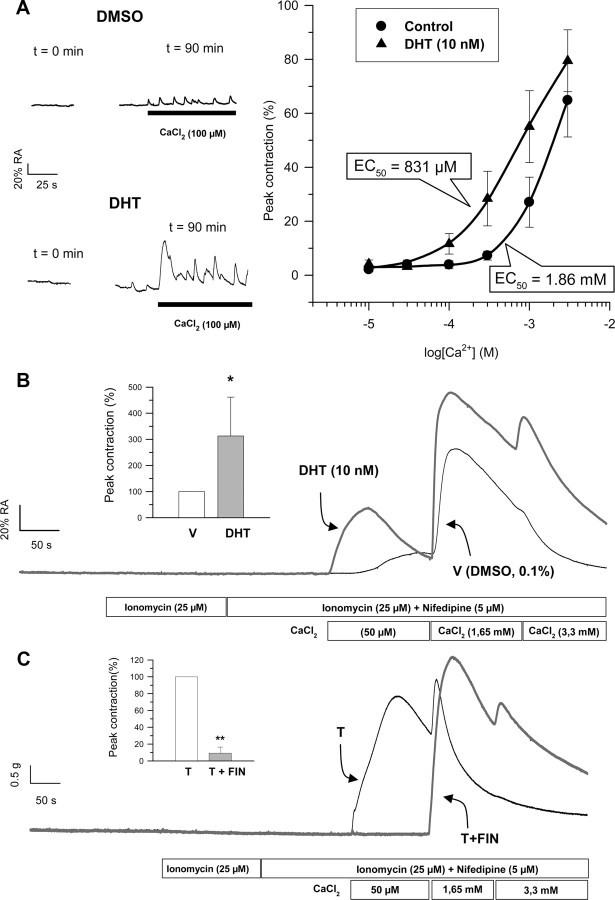
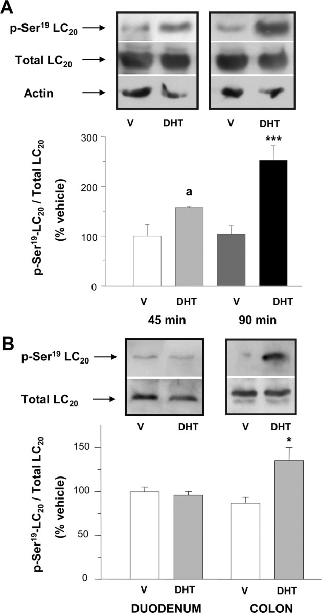
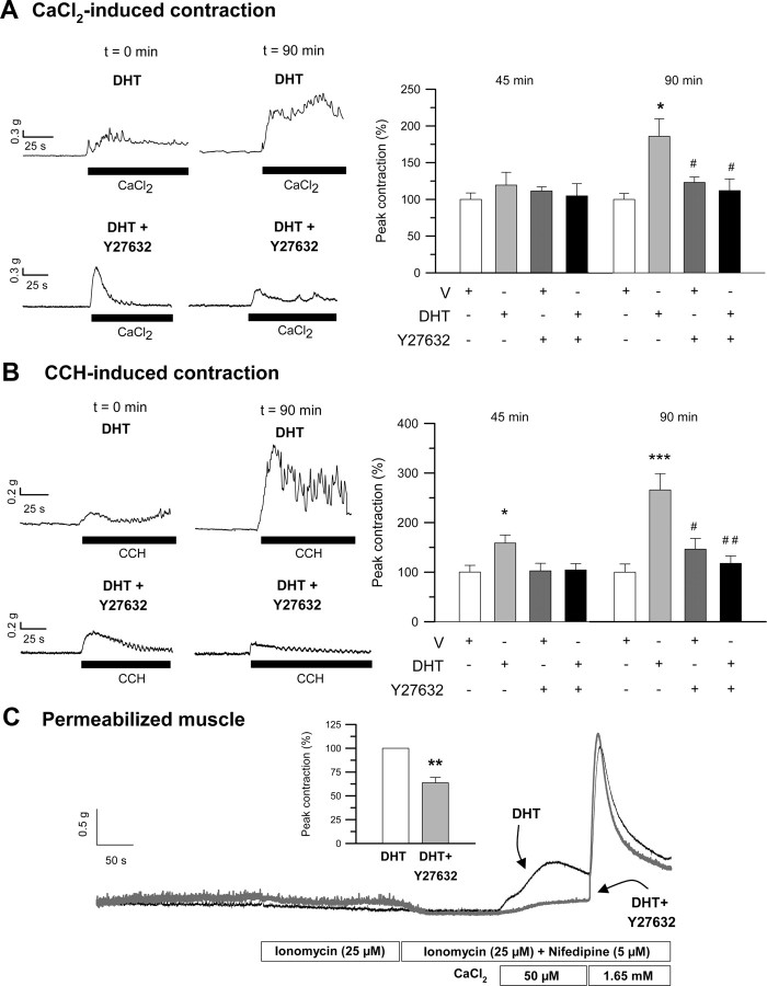
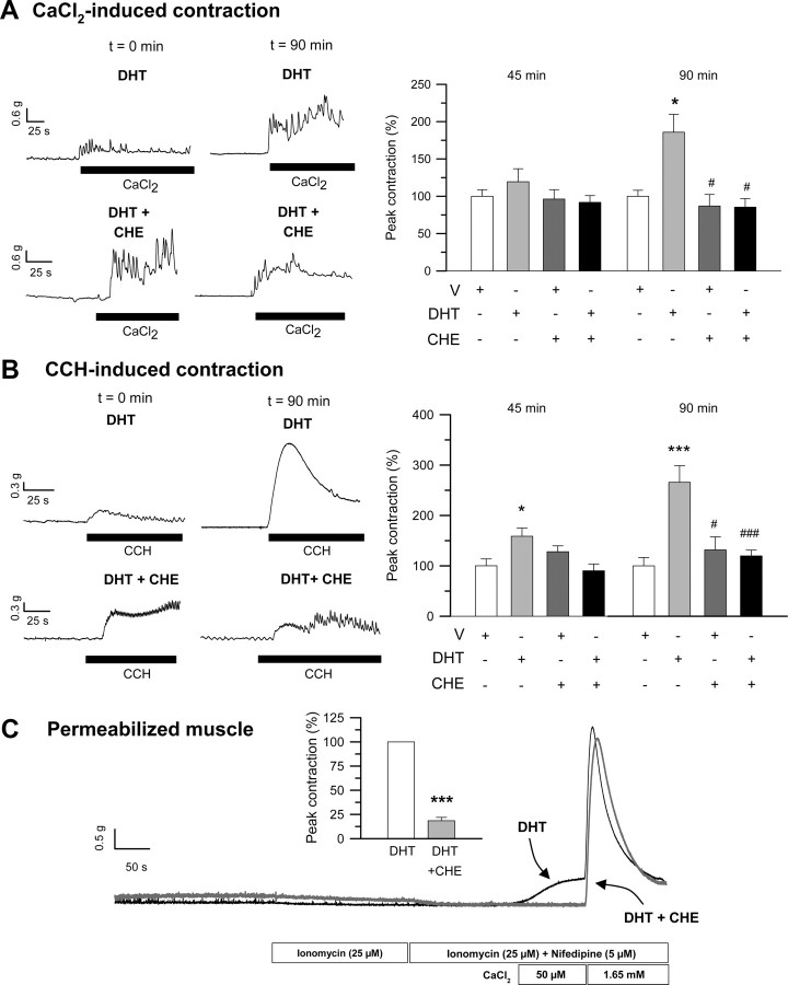
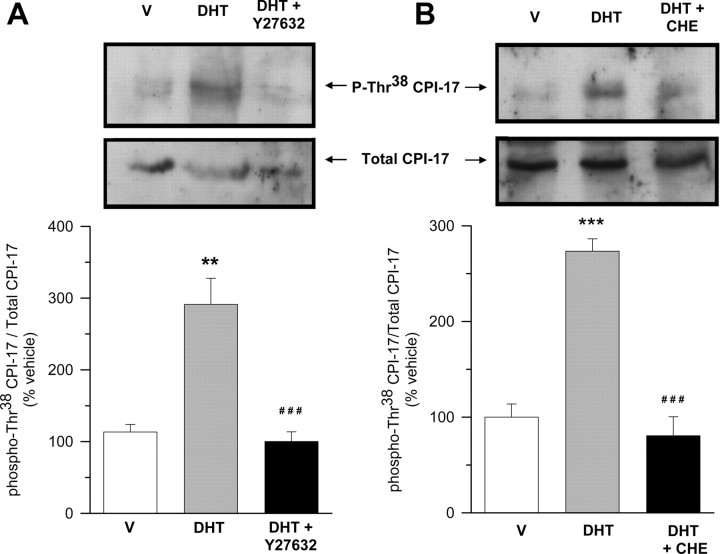
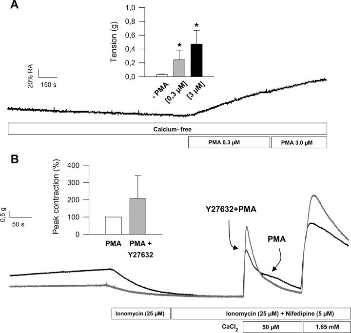
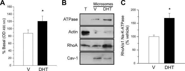
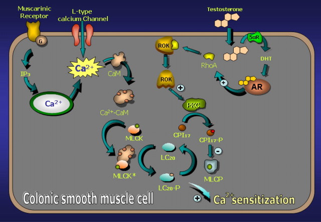
We show that androgens, testosterone and 5alpha-dihydrotestosterone (DHT), acutely (approximately 40 min) provoke the mechanical potentiation of spontaneous and agonist-induced contractile activity in mouse colonic longitudinal smooth muscle. The results using flutamide, finasteride, cycloheximide, and actinomycin D indicate that androgen-induced potentiation is dependent on androgen receptors, requires reduction of testosterone to DHT, and occurs independently of transcriptional and translational events. Using permeabilized colonic smooth muscle preparations, we could demonstrate that mechanical potentiation is entirely due to calcium sensitization of contractile machinery. In addition, DHT (10 nm) increased phosphorylation of both 20-kDa myosin light chain (LC(20)) [regulatory myosin light chain, (MLC)] and CPI-17 (an endogenous inhibitor of MLC phosphatase). Paralleling these findings, inhibition of Rho-associated Rho kinase (ROK) and/or protein kinase C (PKC) with, respectively, Y27632 and chelerythrine, prevented LC(20) phosphorylation and abolished calcium sensitization. In addition, inhibition of ROK prevents CPI-17 phosphorylation, indicating that ROK is located upstream PKC-mediated CPI-17 modulation in the signalling cascade. Additionally, androgens induce a rapid activation of RhoA and its translocation to the plasma membrane to activate ROK. The results demonstrate that androgens induce sensitization of colonic smooth muscle to calcium through activation of ROK, which in turn, activates PKC to induce CPI-17 phosphorylation. Activation of this pathway induces a potent steady stimulation of LC(20) by inhibiting MLC phosphatase and displacing the equilibrium of the regulatory subunit towards its phosphorylated state. This is the first demonstration that colonic smooth muscle is a physiological target for androgen hormones, and that androgens modulate force generation of smooth muscle contractile machinery through nongenomic calcium sensitization pathways.


Figures

Fig. 1.
Fig. 1.
Effects of androgens on contractile responses of colonic and duodenal longitudinal smooth muscles. A and B, Left panels, Representative recordings of the effects of T on contractile responses elicited by CaCl2 (A) and CCH (B). Tissues were exposed to T (10 nm) for 90 min. The contractile responses to CaCl2 and CCH were recorded at the beginning of the experiment (Control) and 90 min after exposure to the hormone. External stimuli were applied after a 5-min period in which tissues were maintained under calcium-free conditions. Right panels, Summary of the effects of preincubation with T (10 nm) and DHT (10 nm) on CaCl2-induced (A) and CCH-induced (B) contractions measured at 45 and 90 min. C, Effects of preincubation of duodenal smooth muscle with T (10 nm) on CaCl2- and CCH-induced contractions. Results in right panels of A and B represent the mean ± sem of 15 different experiments. Statistical significance was assessed by one-way ANOVA, followed by Student’s t test. a, P < 0.1; *, P < 0.05; and ***, P < 0.005 compared with vehicle (V), respectively.
Fig. 2.
Fig. 2.
Androgens alter the frequency spectrum of colonic peristaltic activity. A, Representative recording showing the time course of spontaneous activity in the presence of vehicle DMSO (0.1%) and after application of DHT (10 nm). B, Frequency analyses using the FFT obtained 90 min after incubation with vehicle (red trace) or DHT (blue trace). PSD, Power spectral density. C, Application of the STFT to the contractile signal shown in A. Results are representative of another five different experiments. Color scale (gray) encodes for spectral densities. For details, see Materials and Methods and Results.
Fig. 3.
Fig. 3.
Analysis of AR expression in colon and duodenum. Total protein extracts from colonic (C) and duodenal (D) smooth muscle were electrophoresed on SDS-PAGE and processed for Western blot analysis, using a specific polyclonal antibody to AR. As a control of protein load, membranes were reblotted with an antibody directed to ERα. Molecular messes of the different specific bands recognized by the antibodies are indicated. Results are representative of five experiments.
Fig. 4.
Fig. 4.
Androgens induce Ca2+ sensitization in colonic smooth muscle. A, Calcium dependence curves for control and DHT (10 nm) measured as peak responses to CaCl2 in longitudinal colonic muscle. Tissues were allowed to preincubate with control or DHT for 90 min before application of the corresponding calcium pulse. Traces on the left panel illustrate typical responses to calcium (100 μm) at time 0 and after 90 min preincubation with DHT. B, Typical responses of ionomycin-permeabilized colonic longitudinal muscle to extracellular calcium (50 μm, 1.6 mm, and 3.3 mm) preincubated with vehicle (V) (DMSO, 0.1%, 90 min; trace in black) or DHT (10 nm, 90 min; trace in dark gray). Inset, A summary of the results from another seven experiments. *, P < 0.05 compared with V, respectively. C, Effects of FIN on DHT-induced calcium sensitization. Tissues were allow to preincubated with FIN (10 μm) for 30 min or DMSO (0.1%) before incubation with DHT (10 nm) and then submitted to the permeabilization maneuver and calcium challenges at the concentrations indicated. Inset, A summary of the results from another four experiments. **, P < 0.01 compared with DHT. Recordings (control and experimental) in B and C were obtained in smooth muscle preparations from the same animals.
Fig. 5.
Fig. 5.
Androgens induce the phosphorylation of LC20 in colonic muscle. A, Western blot analyses for total and phosphorylated-Ser19 LC20 (p-Ser19-LC20) in colonic muscle extracts. In each experiment, equivalent colonic segments obtained from the same animal were exposed for 45 or 90 min to either vehicle (V) (DMSO, 0.1%) or DHT (10 nm). Top, Representative assay after immunoblotting with either antiphospho-LC20 or total LC20 antibodies. α-Actin was used as a control of protein load. Bottom, Densitometric values of phospho-LC20 relative to total LC20. Values were normalized to the average of immunosignals obtained with V-treated tissues (V). a and ***, P < 0.1 and P < 0.005 compared with V, respectively. B, Comparison of the effects of preincubation with V (DMSO, 0.1%; 90 min) or DHT (10 nm, 90 min) on the levels of total LC20 and p-Ser19-LC20 between colonic and duodenal segments. Colonic and duodenal segments were obtained from the same animals. Top, Representative assays after immunoblotting with either antiphospho-LC20 or total LC20 antibodies. Bottom, Densitometric values of phospho-LC20 relative to total LC20 and normalized to average immunosignals obtained with V. *, P < 0.05 vs. V-treated tissues. Four assays were performed for each type of experiment and intestinal segment in A and B.
Fig. 6.
Fig. 6.
Involvement of RhoA-associated ROK in the induction of mechanical potentiation and calcium sensitization elicited by androgens. A and B, Left panels, Illustrative recordings, taken at time 0 and 90 min, showing the effect of 45-min preincubation with the specific inhibitor of ROK, Y27632 (10 μm), on DHT-induced (10 nm) potentiation of contractile responses to CaCl2 and CCH. Right panels, Summary of the effects of preincubation with Y27632 on DHT-induced stimulation of CaCl2-triggered (A) and CCH-triggered (B) contractions as measured at 45 and 90 min (n = 6). * and ***, P < 0.05 and P < 0.005 compared with vehicle (V), respectively; # and ##, P < 0.05 and P < 0.01 vs. DHT. C, Effects of Y27632 (10 μm) preincubation (45 min) on DHT-induced calcium sensitization in permeabilized colonic muscle. Inset, A summary of the results, expressed as mean ± sem, from another four experiments. In each experiment, equivalent segments of colonic muscle obtained from the same animal were preexposed to either V or Y27632 (10 μm) for 45 min before incubation with DHT (10 nm, 90 min). **, P < 0.01 vs. DHT.
Fig. 7.
Fig. 7.
Involvement of PKC in the induction of mechanical potentiation and calcium sensitization elicited by androgens. A and B, Left panels, Illustrative recordings, taken at time 0 and 90 min, showing the effect of 20-min preincubation with the generic inhibitor of PKC, CHE (1 μm), on DHT-induced (10 nm) potentiation of contractile responses to CaCl2 and CCH. Right panels, Summary of the effects of preincubation with CHE on DHT-induced stimulation of CaCl2-triggered (A) and CCH-triggered (B) contractions as measured at 45 and 90 min (n = 4). * and ***, P < 0.05 and P < 0.005 compared with vehicle (V), respectively; # and ###, P < 0.05 and P < 0.005 vs. DHT. C, Effects of CHE (1 μm) preincubation (10 min) on DHT-induced calcium sensitization in permeabilized colonic muscle. Inset, A summary of the results, expressed as mean ± sem, from another three experiments. In each experiment, equivalent segments of colonic muscle obtained from the same animal were preexposed to either V or CHE (1 μm) for 45 min before incubation with DHT (10 nm, 90 min). ***, P < 0.005 vs. DHT.
Fig. 8.
Fig. 8.
ROK and PKC are involved in DHT-induced phosphorylation of LC20 in colonic muscle. Western blot analyses for total LC20 and phosphorylated Ser19 LC20 (p-Ser19-LC20) in muscle extracts. In each experiment, equivalent segments from colon obtained from the same animal were incubated in the presence of vehicle (V) (DMSO, 0.1%), DHT (10 nm), or DHT+Y27632 (left panel) or V (DMSO, 0.1%), DHT (10 nm), or DHT+CHE (right panel). Tissues exposed to Y27632 (10 μm) or CHE (1 μm) were allowed to preincubate for 45 or 20 min, respectively, with the inhibitor before application of DHT for additional 90 min. Total lysates were analyzed for changes in LC20 phosphorylation using antiphospho(Ser19)-LC20 antibody, followed by reblotting with an antitotal-LC20 antibody. Top, Representative assays after immunoblotting with antiphospho-LC20 and total LC20 antibodies. Bottom, Densitometric values of phospho-LC20 relative to total LC20 expressed as percentage of V. ###, P < 0.005 vs. V; ** and ***, P < 0.01 and P < 0.005 vs. DHT-treated tissues. Four assays were performed under each condition.
Fig. 9.
Fig. 9.
ROK and PKC are involved in DHT-induced phosphorylation of CPI-17 in colonic muscle. Western blot analyses for total CPI-17 and phosphorylated Thr38 CPI-17 (p-Thr38-CPI-17) in colonic muscle extracts. In each experiment, equivalent segments from colon obtained from the same animal were incubated in the presence of vehicle (V) (DMSO, 0.1%), DHT (10 nm), or DHT+Y27632 (left panel) or V (DMSO, 0.1%), DHT (10 nm), or DHT+CHE (right panel). Tissues exposed to Y27632 (10 μm) or CHE (1 μm) were allowed to preincubate for 45 or 20 min, respectively, with the inhibitor before application of DHT for additional 90 min. Total lysates were analyzed for changes in CPI-17 phosphorylation using antiphospho(Thr38)-CPI-17 antibody, followed by reblotting with an antitotal-CPI-17 antibody. Top, Representative assays after immunoblotting with antiphospho-CPI-17 and total CPI-17 antibodies. Bottom, Densitometric values of phospho-CPI-17 relative to total CPI-17 expressed as percentage of V. ** and ***, P < 0.01 and P < 0.005 vs. V; ###, P < 0.005 vs. DHT-treated tissues. Four assays were performed under each condition.
Fig. 10.
Fig. 10.
A, PMA induces Ca2+ sensitization in colonic smooth muscle. Illustrative response of colonic longitudinal muscle to extracellular increasing concentrations of PMA (0.3–3 μm) under extracellular calcium-free conditions. Inset, A summary of the results from four different dose-response experiments. *, P < 0.05 vs. vehicle (DMSO, 0.1%). B, Inhibition of ROK does not prevent PMA-induced Ca2+ sensitization in colonic smooth muscle. Illustrative response of ionomycin-permeabilized colonic longitudinal muscle to extracellular calcium pulses. Tissues were preincubated with Y27632 (10 μm, 45 min; black trace) or vehicle (dark gray trace) before being exposed to PMA (0.3 μm, 10 min) and submitted to the permeabilization maneuver and calcium challenges at the concentrations indicated. Inset, A summary of the results from another three experiments. Recordings (PMA and PMA + Y27632) were obtained from colonic smooth muscle preparations from the same animal.
Fig. 11.
Fig. 11.
Androgens elicit RhoA activation and translocation to the plasma membrane. A, Determination of activated RhoA (GTP·RhoA) in colonic muscle preparations preexposed to vehicle (V) (DMSO, 0.1%) or DHT (10 nm, 20 min). *, P < 0.05 compared with V. B, Representative assay after immunoblotting with anti-RhoA and plasma membrane markers (α1 Na+/K+-ATPase and caveolin-1) in total lysates (T) and microsomal fractions from colonic muscle. The cytosolic marker α-actin was used as a control of microsomes purity. Colonic tissues were exposed to V (DMSO 0.1%) or DHT (10 nm). C, Densitometric values of RhoA relative to α1 Na+/K+-ATPase expressed as percentage of V. *, P < 0.05 vs. V-treated tissues. Four assays were performed under each condition in all experiments.
Fig. 12.
Fig. 12.
Proposed cellular model illustrating the mechanisms of Ca2+ sensitization in mouse colonic muscle induced by androgens. For details see Discussion. CaM, Calmodulin.

Similar articles

Cited by

References

    1. Foradori CD, Weiser MJ, Handa RJ2008. Non-genomic actions of androgens. Front Neuroendocrinol 29:169–181 - PMC - PubMed
    1. Michels G, Hoppe UC2008. Rapid actions of androgens. Front Neuroendocrinol 29:182–198 - PubMed
    1. Lieberherr M, Grosse B1994. Androgens increase intracellular calcium concentration and inositol 1,4,5-trisphosphate and diacylglycerol formation via a pertussis toxin- sensitive G-protein. J Biol Chem 269:7217–7223 - PubMed
    1. Guo Z, Benten WP, Krücken J, Wunderlich F2002. Nongenomic testosterone calcium signaling. Genotropic actions in androgen receptor-free macrophages. J Biol Chem 277:29600–29607 - PubMed
    1. Benten WP, Lieberherr M, Giese G, Wrehlke C, Stamm O, Sekeris CE, Mossmann H, Wunderlich F1999. Functional testosterone receptors in plasma membranes of T cells. FASEB J 13:123–133 - PubMed

Publication types

MeSH terms