. 2010 Mar;95(3):432-9.
doi: 10.3324/haematol.2009.010991.

The inducible T-cell co-stimulator molecule is expressed on subsets of T cells and is a new marker of lymphomas of T follicular helper cell-derivation

Affiliations

The inducible T-cell co-stimulator molecule is expressed on subsets of T cells and is a new marker of lymphomas of T follicular helper cell-derivation

Teresa Marafioti et al. Haematologica. 2010 Mar.

Abstract

Background: T follicular helper (T(FH)) cells reside in the light zone of germinal centers and are considered the cell of origin of angioimmunoblastic T-cell lymphoma. Recently, CXCL13, PD-1 and SAP were described as useful markers for T(FH) cells and angioimmunoblastic T-cell lymphoma but also reported in some peripheral T-cell lymphomas, not otherwise specified.

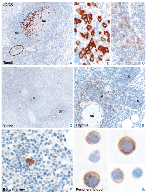
Design and methods: In the present study the expression pattern of ICOS protein was investigated by immunohistochemistry-based techniques in routine sections of normal lymphoid tissues and 633 human lymphomas.

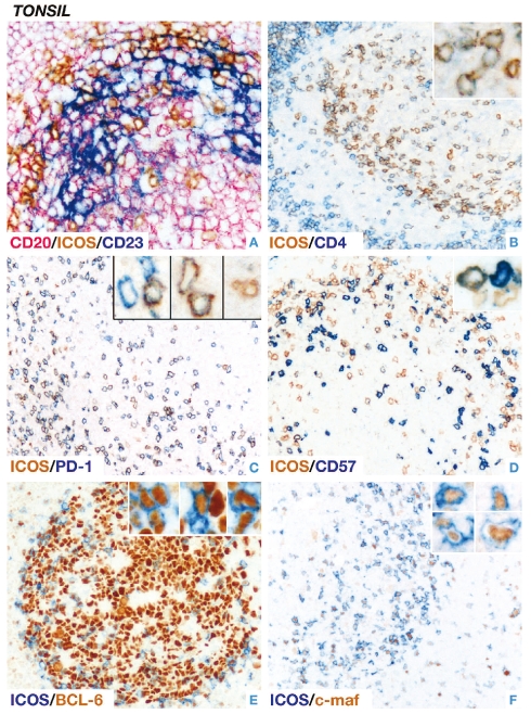
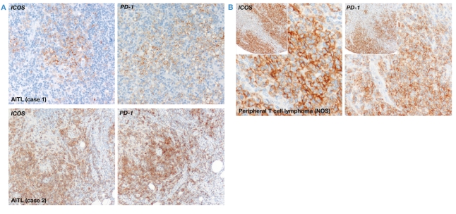
Results: Cells strongly positive for ICOS were restricted to the light zone of germinal centers and co-expressed T(FH)-associated molecules. In addition, weak to moderate ICOS expression was observed in a small proportion of FOXP3-positive cells. In lymphomas, ICOS expression was confined to angioimmunoblastic T-cell lymphoma (85/86), peripheral T-cell lymphomas of follicular variant (18/18) and a proportion of peripheral T-cell lymphomas, not otherwise specified (24/56) that also expressed other T(FH)-associated molecules.

Conclusions: ICOS is a useful molecule for identifying T(FH) cells and its restricted expression to angioimmunoblastic T-cell lymphoma and a proportion of peripheral T-cell lymphomas, not otherwise specified (showing a T(FH)-like profile) suggests its inclusion in the antibody panel for diagnosing T(FH)-derived lymphomas. Our findings provide further evidence that the histological spectrum of T(FH)-derived lymphomas is broader than previously assumed.


Figures

Figure 1.
Figure 1.
Immunostaining of ICOS in normal human lymphoid tissues. (A) In tonsil, strong ICOS labeling is seen in a population of lymphoid cells lying at the periphery of the light zone of a germinal center (boxed area). In the interfollicular area, many T-cells (circled area) weakly express ICOS (GC: germinal center; MC: mantle cells) (X200). (B) The image illustrates, at higher magnification, strong membrane and cytoplasmic associated ICOS staining of a group of intra-germinal center cells (X600). (C) T-cells in the interfollicular area, shown at higher magnification, are weakly ICOS-positive (X400). (D) In the spleen rare scattered ICOS-positive cells are usually seen in both the red and white pulp. Note the presence of two germinal centers containing many ICOS-positive cells (X100). (E) Many cells in the thymic medulla are ICOS-positive and only rare scattered ICOS-positive cells can be seen in the thymic cortex (M: medulla; C: cortex; HC: Hassal’s corpuscle) (X200). Rare ICOS-positive cells can also be seen in (F) a bone marrow trephine section (X600) and (G) cytospin preparations of peripheral blood from healthy donors (X600). Staining was performed by the immunoperoxidase technique in paraffin-embedded tissue sections or fresh peripheral blood samples using a polyclonal ICOS antibody and hematoxylin counterstain.
Figure 2
Figure 2
(left). Immunolabeling of ICOS-positive cells in normal human tonsil. (A) Triple immunoenzymatic labeling shows that ICOS-positive cells (brown) are in close contact with CD23-positive follicular dendritic cells (blue) and CD20-positive B-cells (pink) (X400). Double immunoenzymatic labeling demonstrates that (B) the great majority of ICOS-positive (brown) intra-germinal center cells express the T helper cell marker CD4 (blue) (X200) and (C) the TFH-associated molecule PD-1 (blue) (X200). (D) Only a subset of ICOS-positive (brown) intra-germinal center cells co-express the CD57 antigen (blue) (X200). (E–F) The transcription factors BCL-6 (X400) and c-maf (brown, nuclear) (X200) are found in a proportion of ICOS-positive intra-germinal center cells. The insets show examples of the respective staining at higher magnification (all X600). All staining was performed on paraffin-embedded tissue sections and hematoxylin counterstain was omitted.
Figure 3.
Figure 3.
Immunolabeling of ICOS-positive cells in normal human lymphoid tissues. (A) Double immunoenzymatic labeling of tonsil shows, in a germinal center (GC), rare ICOS-positive (brown) cells co-expressing the regulatory T-cell associated transcription factor FOXP3 (blue, nuclear) (x400). (B) In contrast, a higher number of ICOS/FOXP3 double-positive cells are observed in the interfollicular area (X400). (A–B) The insets show examples of double-positive cells at a higher magnification (X600). (C) Triple staining for ICOS (blue), FOXP3 (brown) and the B-cell associated molecule CD79a (pink) in tissue sections of thymus reveals the presence of numerous ICOS/FOXP3 double-positive, CD79a-negative cells (as also shown at higher magnification in the inset, X600) which are mainly localized in the medulla (X200). Double immunoenzymatic labeling in tonsil (D–E) shows that rare ICOS-positive cells (brown) co-express the cytotoxic T-cell molecules CD8 (blue) (X600) and granzyme B (blue) (X600). The arrows indicate double-positive cells; (F) rare CD30-positive (blue) cells (mainly localized in the interfollicular area) co-express ICOS (brown) (X200) as also illustrated at higher magnification in the inset (X600). All staining was performed on paraffin-embedded tissue sections and hematoxylin counterstain was omitted.
Figure 4.
Figure 4.
Immunostaining of ICOS and PD-1 in human T-cell lymphomas. (A) Tumor cells of two cases of angioimmunoblastic T-cell lymphoma (AITL) express the co-stimulatory molecule ICOS and the TFH-associated marker PD-1 (X200). (B) Tumor cells in a case of peripheral T-cell lymphoma not otherwise specified (NOS) show strong expression of ICOS and PD-1 (X400). The insets illustrate, at lower magnification (X100) the staining pattern of ICOS and PD-1. Staining of paraffin-embedded whole or tissue-array sections was performed by the immunoperoxidase technique and counterstained with hematoxylin.

Comment in

Similar articles

Cited by

References

    1. Attygalle A, Al-Jehani R, Diss TC, Munson P, Liu H, Du MQ, et al. Neoplastic T cells in angioimmunoblastic T-cell lymphoma express CD10. Blood. 2002;99(5):627–33. - PubMed
    1. Dogan A, Attygalle AD, Kyriakou C. Angioimmunoblastic T-cell lymphoma. Br J Haematol. 2003;121(5):681–91. - PubMed
    1. Yuan CM, Vergilio JA, Zhao XF, Smith TK, Harris NL, Bagg A. CD10 and BCL6 expression in the diagnosis of angioimmunoblastic T-cell lymphoma: utility of detecting CD10+ T cells by flow cytometry. Human Pathol. 2005;36(7):784–91. - PubMed
    1. Chtanova T, Tangye SG, Newton R, Frank N, Hodge MR, Rolph MS, et al. T follicular helper cells express a distinctive transcriptional profile, reflecting their role as non-Th1/Th2 effector cells that provide help for B cells. J Immunol. 2004;173(1):68–78. - PubMed
    1. Kim CH, Lim HW, Kim JR, Rott L, Hillsamer P, Butcher EC. Unique gene expression program of human germinal center T helper cells. Blood. 2004;104(7):1952–60. - PubMed

Publication types

MeSH terms

Substances