. 2010 Aug;9(8):1171-83.
doi: 10.1128/EC.00329-09. Epub 2010 Mar 5.

Nuclear dynamics, mitosis, and the cytoskeleton during the early stages of colony initiation in Neurospora crassa

Affiliations

Nuclear dynamics, mitosis, and the cytoskeleton during the early stages of colony initiation in Neurospora crassa

M Gabriela Roca et al. Eukaryot Cell. 2010 Aug.

Abstract

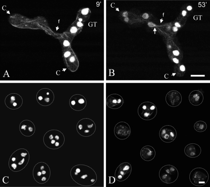
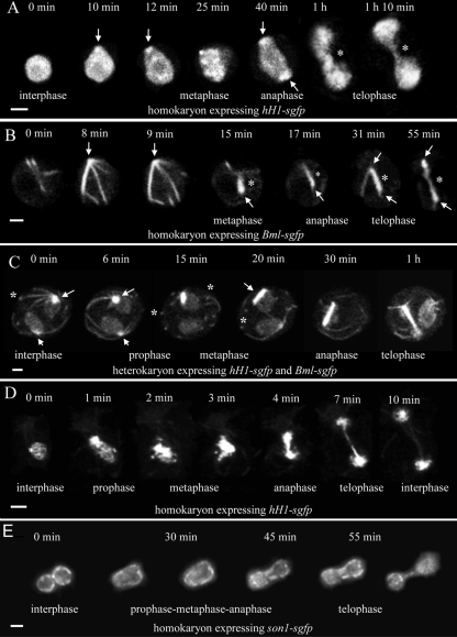
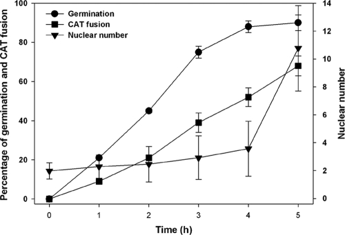
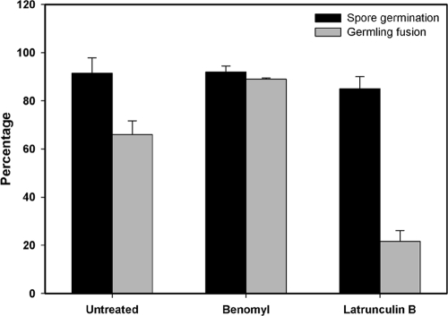
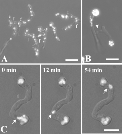
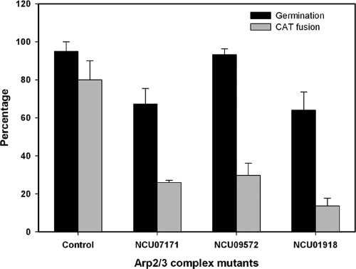
Neurospora crassa macroconidia form germ tubes that are involved in colony establishment and conidial anastomosis tubes (CATs) that fuse to form interconnected networks of conidial germlings. Nuclear and cytoskeletal behaviors were analyzed in macroconidia, germ tubes, and CATs in strains that expressed fluorescently labeled proteins. Heterokaryons formed by CAT fusion provided a rapid method for the imaging of multiple labeled fusion proteins and minimized the potential risk of overexpression artifacts. Mitosis occurred more slowly in nongerminated macroconidia (1.0 to 1.5 h) than in germ tubes (16 to 20 min). The nucleoporin SON-1 was not released from the nuclear envelope during mitosis, which suggests that N. crassa exhibits a form of "closed mitosis." During CAT homing, nuclei did not enter CATs, and mitosis was arrested. Benomyl treatment showed that CAT induction, homing, fusion, as well as nuclear migration through fused CATs do not require microtubules or mitosis. Three ropy mutants (ro-1, ro-3, and ro-11) defective in the dynein/dynactin microtubule motor were impaired in nuclear positioning, but nuclei still migrated through fused CATs. Latrunculin B treatment, imaging of F-actin in living cells using Lifeact-red fluorescent protein (RFP), and analysis of mutants defective in the Arp2/3 complex demonstrated that actin plays important roles in CAT fusion.


Figures

Fig. 1.
Fig. 1.
Timing of fluorescent protein labeling of heterokaryons formed by CAT fusion. (A and B) Fused conidial germlings (one labeled with H1-GFP and the other labeled with β-tubulin-GFP [BML-GFP]), one 9 min (A) and the other 53 min (B) after fusion. At the 9-min time point, BML-GFP has become incorporated into microtubules derived from the left-hand germling with nuclei carrying the hH1-sgfp gene, while HI-GFP has not yet labeled the nuclei in the germling on the right-hand side with nuclei carrying the bml-sgfp gene. Microtubules passed through the fused CATs (f) before nuclei did. The location of the spindle pole body (arrow) with associated microtubules is shown at 53 min. C, conidium; GT, germ tube. Bar, 10 μm. (C) Conidia from a homokaryotic H1-GFP strain. Each conidium has several nuclei. (D) Conidia from a heterokaryotic H1-GFP plus β-tubulin-GFP strain visualized by confocal microscopy. Variable levels of GFP expression were observed for different conidia. Bar, 5 μm.
Fig. 2.
Fig. 2.
Time courses of mitosis in multinucleate nongerminated conidia (A to C) and germ tubes (D and E). The interphase, prophase, metaphase, anaphase, and telophase stages of mitosis have been indicated where they can be clearly recognized. (A) Mitosis of a conidial nucleus with H1-GFP labeling. A bright fluorescent nuclear region (arrows) was often evident where a spindle pole body (SPB) should be located. The segregating H1-containing chromatin was connected by a “chromatin bridge” (*) from late anaphase to telophase. Bar, 2 μm. (B) Mitosis of a conidial nucleus labeled with β-tubulin-GFP. Microtubules were formed from a focal point presumed to be the SPB (arrows). During late prophase/early metaphase, microtubules bundled to form a compact spindle (*), which is evident from metaphase to telophase. Bar, 2 μm. (C) Mitosis in a heterokaryotic conidium in which two nuclei labeled with H1-GFP and β-tubulin-GFP were dividing asynchronously (only one nucleus divided during this time course). Astral microtubules are formed from the single SPBs (arrows) in nuclei during both interphase and early prophase. These microtubules often possessed a bright focal point where they were in contact with the plasma membrane (*). SPB duplication occurred during prophase (at 15 min). The spindle developed as a compact bundle of microtubules (15 min to 1 h). During this time, H1-GFP was associated with the spindle and by telophase had segregated to both SPBs (the chromatin bridge is masked by the BML-GFP in this image). Astral ray microtubules can be visualized at the 1-h time point. Bar, 4 μm. (D) Mitosis in a germ tube nucleus labeled with H1-GFP. In this sequence, the expression level of H1-GFP was much lower than that in C. Bar, 2 μm. (E) Germ tube nucleus labeled with SON-1-GFP to show the nuclear pores in the nuclear envelope. Telophase was easily observed because the bridge formed between the nuclei daughters. Projections of 12 deconvolved images are shown. Bar, 2 μm.
Fig. 3.
Fig. 3.
Rates of conidial germination and CAT fusion and the number of nuclei in conidia/conidial germlings during the first 5 h of incubation. The nuclear number significantly increased only after 4 h, when germination was near maximal and over 50% of the conidial germlings had undergone CAT fusion.
Fig. 4.
Fig. 4.
Cell cycle arrest during CAT homing. (A) CATs formed from germ tubes before (0 min) and after (5 and 7 min) fusion visualized by confocal microscopy. The upper conidial germling was labeled with H1-GFP, while the lower germling was labeled with β-tubulin-GFP. Bar, 5 μm. (B) Percentages of nuclei undergoing mitosis in ungerminated macroconidia (0 to 3 h), in macroconidia possessing only germ tubes (2 to 4 h), in macroconidia or macroconidial germlings possessing CATs (3 to 4 h), and in macroconidia or macroconidial germlings that have undergone CAT fusion (4 to 6 h). The time periods selected for the analysis of mitosis in these four different cell types were those in which the production of the individual cell types was maximal. Mitotic nuclei were identified by the presence of thick microtubule bundles in spindles labeled with BML-GFP.
Fig. 5.
Fig. 5.
Microtubular and nuclear organization and dynamics visualized by confocal and bright-field microscopy. (A) Two adjacent optical sections (0.5 μm apart) of a heterokaryotic conidium and germ tube showing five nuclei (n) after labeling with H1-GFP and β-tubulin-GFP, visualized by confocal microscopy. These two optical sections were selected from a continuous z stack of 20 optical sections captured in 0.5-μm steps. Arrows indicate the locations of spindle pole bodies with associated microtubules. Nuclei appear to be linked via microtubules. A large vacuole (v) seems to have pushed two of the nuclei to the periphery of the conidium. C, conidium; GT, germ tube. Bar, 5 μm. (B) Lack of inhibition of CAT fusion and homing by treatment with the microtubule-depolymerizing drug benomyl (40 μg ml−1). Two hours after treatment, the microtubules were reduced to punctate spots. Bar, 10 μm. (C) Nuclei labeled with H1-GFP migrate through fused CATs visualized by combined confocal and bright-field microscopy. (D) Benomyl treatment prevents the formation of long germ tubes. An H1-GFP-labeled strain was incubated with benomyl (35 μg ml−1) for 6 h visualized by combined wide-field fluorescence and differential interference contrast (DIC) microscopy. Small buds (arrow) may represent the initiation of germ tube formation, but these buds did not elongate further. Bar, 5 μm.
Fig. 6.
Fig. 6.
Quantification of effects of the antimicrotubule drug benomyl (40 μg ml−1) and the antiactin drug latrunculin B (40 μg ml−1) on macroconidial germination and CAT fusion. Conidial germination represents the percentage of macroconidia possessing germ tubes and/or CATs. CAT fusion was reduced greatly with the sublethal latrunculin B treatment.
Fig. 7.
Fig. 7.
Nuclear distribution and behavior in the wild type and ropy (dynein/dynactin) mutants visualized by combined wide-field, bright-field, and fluorescence microscopy. Nuclei were labeled with H1-GFP. (A) Network of fused wild-type germlings showing a more-or-less random distribution of nuclei. (B) ro-1 (dynein heavy chain) mutant showing CATs derived from germ tubes homing toward each other (arrow). The nuclei are concentrated in or close to the conidia. (C) Δro-11 (dynactin subunit) mutant showing nuclear migration (arrow) through fused CATs (f) after incubation for 7 h. f, points of CAT fusion. Bars, 15 μm.
Fig. 8.
Fig. 8.
Greater inhibition of CAT fusion than conidial germination in deletion mutants defective in different protein subunits within ARP2/3 protein complex.
Fig. 9.
Fig. 9.
F-actin dynamics during CAT-mediated cell fusion are unaffected by benomyl treatment. Shown are pairs of Lifeact-RFP-expressing conidia of N. crassa during pre- and postfusion stages. (A) In the untreated control, fluorescence intensities of actin cables and patches are concentrated at the CAT tips during homing and peak as cell-to-cell contact is established. (B) After cytoplasmic continuity has been achieved by fusion pore formation, the fluorescence intensity has decreased, and the remaining patches are dispersed. (C and D) In the presence of 3 μg/ml benomyl, germ tubes are unable to become elongated, and the macroconidia show extended isotropic growth and become swollen. The localization and abundance of actin cables and patches remained essentially unchanged after benomyl treatment. Arrows mark sites of CAT contact/fusion. Scale bars, 5 μm.
Fig. 10.
Fig. 10.
Summary of the main features of the different stages of mitosis in a nongerminated macroconidium of N. crassa. During interphase, nuclei (dark green) are very mobile, rotate, and are rounded or pear shaped. When rounded, they are ∼2 μm in diameter. Astral ray microtubules exhibiting pronounced dynamic instability (red lines) form from a single spindle pole body (SPB) (yellow spot) on the surface of the nucleus. During prophase, nuclei are very mobile, rotate, and are rounded or pear shaped. When rounded, they are <4 μm in diameter. The SPB duplicates, and the two daughter SPBs start to move apart. Astral ray microtubules continue to exhibit pronounced dynamic instability. Condensed chromatin starts to appear toward the end of prophase. During metaphase, nuclei are less mobile, rotate less, and have an irregular shape. Astral microtubules seem to disappear. Condensed chromatin (darker green regions within the nucleoplasm) becomes prominent. A spindle composed of a bundle of microtubules (thick red line) grows in length between the SPBs that continue to move apart. During anaphase, future daughter nuclei start to move apart and start to become mobile and rotate again. Short astral microtubules start to form again. The SPBs are located at the two poles of the dividing nuclei with a long spindle bundle between them. During telophase, constriction between future daughter nuclei becomes prominent. Nuclear mobility and rotations are similar to those in anaphase. A long spindle composed of a microtubule bundle (termed “telophase bundle”) has chromatin (the so-called “chromatin bridge”) associated with it. This is visible until the daughter nuclei separate during nucleokinesis. During interphase, daughter nuclei with variable morphologies have been formed (see above).

Similar articles

Cited by

References

    1. Aist J. R. 2002. Mitosis and motor proteins in the filamentous ascomycete, Nectria haematococca, and some related fungi. Int. Rev. Cytol. 212:239–263 - PubMed
    1. Aist J. R., Morris N. R. 1999. Mitosis in filamentous fungi: how we got where we are. Fungal Genet. Biol. 27:1–25 - PubMed
    1. Araujo-Bazán L., Peñalva M. A., Espeso E. A. 2008. Preferential localization of the endocytic internalization machinery to hyphal tips underlies polarization of the actin cytoskeleton in Aspergillus nidulans. Mol. Microbiol. 67:891–905 - PubMed
    1. Araujo-Palomares C. L., Castro-Longoria E., Riquelme M. 2007. Ontogeny of the Spitzenkörper in germlings of Neurospora crassa. Fungal Genet. Biol. 44:492–503 - PubMed
    1. Asakura T., Sasaki T., Nagano F., Satoh A., Obaishi H., Nishioka H., Imamura H., Hotta K., Tanaka K., Nakanishi H., Takai Y. 1998. Isolation and characterization of a novel actin filament-binding protein from Saccharomyces cerevisiae. Oncogene 16:121–130 - PubMed

Publication types

MeSH terms