. 2010 Jul;51(7):3379-86.
doi: 10.1167/iovs.09-4853. Epub 2010 Mar 5.

Identification of novel substrates for the serine protease HTRA1 in the human RPE secretome

Affiliations

Identification of novel substrates for the serine protease HTRA1 in the human RPE secretome

Eunkyung An et al. Invest Ophthalmol Vis Sci. 2010 Jul.

Abstract

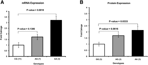
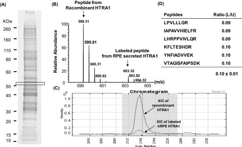
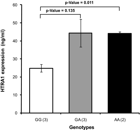
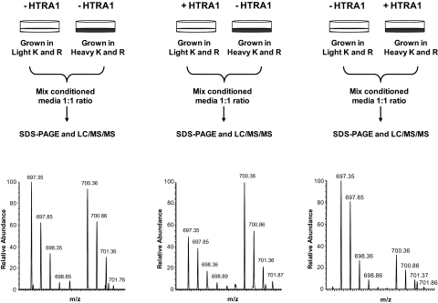
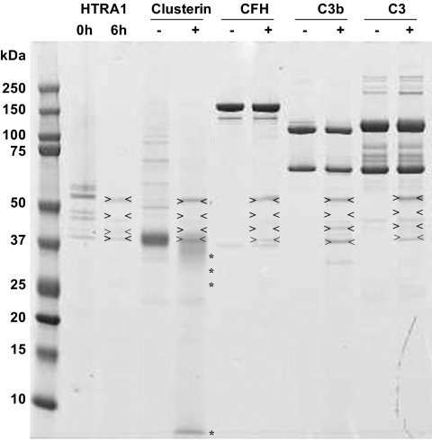
PURPOSE. To define the role of the serine protease HTRA1 in age-related macular degeneration (AMD) by examining its expression level and identifying its potential substrates in the context of primary RPE cell extracellular milieu. METHODS. Primary RPE cell cultures were established from human donor eyes and screened for CFH, ARMS2, and HTRA1 risk genotypes by using an allele-discrimination assay. HTRA1 expression in genotyped RPE cells was determined by using real-time PCR and quantitative proteomics. Potential HTRA1 substrates were identified by incubating RPE-conditioned medium with or without human recombinant HTRA1. Selectively cleaved proteins were quantified by using the differential stable isotope labeling by amino acids in cell culture (SILAC) strategy. RESULTS. HTRA1 mRNA levels were threefold higher in primary RPE cells homozygous for the HTRA1 promoter risk allele than in RPE cells with the wild-type allele, which translated into a twofold increase in HTRA1 secretion by RPE cells with the risk genotype. A total of 196 extracellular proteins were identified in the RPE secretome, and only 8 were found to be selectively cleaved by the human recombinant HTRA1. These include fibromodulin with 90% cleavage, clusterin (50%), ADAM9 (54%), vitronectin (54%), and alpha2-macroglobulin (55%), as well as some cell surface proteins including talin-1 (21%), fascin (40%), and chloride intracellular channel protein 1 (51%). CONCLUSIONS. Recombinant HTRA1 cleaves RPE-secreted proteins involved in regulation of the complement pathway (clusterin, vitronectin, and fibromodulin) and of amyloid deposition (clusterin, alpha2-macroglobulin, and ADAM9). These findings suggest a link between HTRA1, complement regulation, and amyloid deposition in AMD pathogenesis.


Figures

Figure 1.
Figure 1.
Structure of the full-length human HTRA1 protein. Boxes represent protein domains and numbers indicate amino acid positions. SP, signal peptide domain (1-22) cleaved in mature secreted protein; IGFBP, insulin growth factor–binding protein domain (33-100); Kazal-like: Kazal-like domain, with a structure similar to that of Kazal-type serine protease inhibitors. This domain may be responsible for the inhibition of autocleavage of HTRA1 (101-155). Serine protease domain: contains HDS, a catalytic triad (204-364); PDZ, postsynaptic density protein (psd95), drosophila disc large tumor suppressor (dlga), and zonula occludens-1 protein (zo-1) domain (365-467) recognize a broad range of hydrophobic polypeptides in the HTRA1 substrate.
Figure 2.
Figure 2.
Expression levels of HTRA1 mRNA and protein in human RPE cell cultures derived from donors with wild-type and at-risk genotypes for the HTRA1 promoter region. (A) qRT PCR of HTRA1 mRNA levels in primary RPE cultures. Samples were prepared from RPE cultures with wild-type (n = 11), heterozygous (n = 7), and homozygous (n = 3) genotypes for the HTRA1 promoter risk allele (G is the wild-type allele and A is the risk allele). Total RNA was isolated from cultured RPE cells. cDNA was prepared by reverse transcription. qRT-PCR was performed with forward and reverse primers that recognize exons 6 and 7 of the HTRA1 gene. HTRA1 mRNA transcript levels were measured. All values were normalized to wild-type GG genotype. (B) Western blot analysis of HTRA1 protein levels in the conditioned media of RPE cultures. RPE cells with wild-type genotype GG (n = 3), at-risk heterozygous genotype AG (n = 3), and at-risk homozygous genotype AA (n = 3) were grown in serum-containing medium. After the cells reached confluence, the medium was replaced with serum-free medium for 24 hours. Media were collected, and an equal amount of proteins (10 μg) from each sample was concentrated and resolved by SDS-PAGE for Western blot analysis. HTRA1 band intensities were normalized to total loaded proteins. Error bars, SE.
Figure 3.
Figure 3.
Stable-isotope–labeled standard for absolute quantification of HTRA1 protein. Conditioned medium collected from a fully labeled RPE culture grown in medium containing 13C6-Arg, 13C6,15N2-Lys was spiked with human recombinant unlabeled HTRA1 at a final concentration of 200 ng/mL. Samples were run on SDS-PAGE, bands were sliced and digested by trypsin, and the resulting peptides were analyzed by LC-MS/SM. (A) SDS-PAGE of total secreted proteins collected from a 24-hour RPE culture grown in SILAC-labeled, serum-free medium. As expected, HTRA1 protein was identified with six different peptides ∼50 kDa located in the boxed area. (B) Example of a mass spectrum for the YNFIADVVEK peptide of HTRA1. This peptide was detected as a pair of unlabeled (599.31 m/z) and labeled peptide (603.31 m/z), both as double-charged ions. Both these peptides were sequenced, and the difference in mass of 8 Da (4 Da for the double-charged ions) agrees with a Lys residue at the C-terminal. (C) The intensity ratio of heavy to light peak was determined from an XIC for unlabeled and labeled peptide pairs using Census software and reflects the ratio of endogenous RPE HTRA1 to the recombinant HTRA1. (D) A total of six peptide pairs were detected for HTRA1, and the ratios between unlabeled and labeled peptide pairs was measured. Since the amount of recombinant HTRA1 added to the conditioned medium is known, it allowed us to calculate that 10-cm culture dish containing 5 × 10⁶ RPE cells secrete approximately 20 ± 1.49 ng/mL of HTRA1.
Figure 4.
Figure 4.
Effect of the HTRA1 promoter genotype on HTRA1 expression in RPE cultures. Stable-isotope–labeled HTRA1 (20 ng/mL) was used as a standard to spike conditioned media collected from fully confluent RPE cultures with the homozygous wild-type genotype GG (n = 3), the heterozygous at-risk genotype GA (n = 3), and homozygous at-risk genotype AA (n = 2) for the HTRA1 promoter. Quantities of secreted HTRA1 were calculated from intensity ratios of light and heavy peptide pairs detected from the unlabeled RPE-secreted HTRA1 and the stable-isotope–labeled HTRA1 standard. The number of unique peptides used to determine HTRA1 in each genotype group was as follows: eight peptides for the GG variant group, four for the GA variant, and four for the AA variant. Error bars, SE between replicate samples analyzed for each genotype.
Figure 5.
Figure 5.
Differential SILAC strategy for screening for HTRA1 substrates in RPE-conditioned medium. Top: SILAC-labeled or unlabeled conditioned media were incubated with (+HTRA1) or without (−HTRA1) for 20 hours at 37°C. Treated and untreated conditioned media were then mixed at 1:1 ratio and processed for SDS-PAGE and LC-MS/MS. Clusterin was identified by 15 labeled and unlabeled peptide pairs in the RPE secretome. Bottom: mass spectra for one of the clusterin peptide detected as a pair of SILAC-labeled and unlabeled peptides. The intensity ratio of these peaks was equal to 1 when both samples were untreated with HTRA1 (left), but decreased to 0.5 in treated versus untreated sample pairs (center and right), clearly indicating that clusterin was degraded by HTRA1.
Figure 6.
Figure 6.
Clusterin was a specific substrate for HTRA1. The results of a shotgun proteome profiling analysis suggested that clusterin is selectively cleaved by HTRA1. To validate this finding, three recombinant proteins regulating the complement pathway, including human recombinant clusterin and purified human CFH, C3, and C3b were incubated with human recombinant HTRA1. Each protein (2 μg) was incubated with (+) and without (−) recombinant HTRA1 (0.08 μg) for 6 hours at 37°C (enzyme to protein ratio, 1:25, wt/wt) in 4 μL solution of 50 mM Tris-HCl (pH 7.5), 150 mM NaCl, and 5 mM CaCl2. Whereas clusterin was significantly degraded by HTRA1 (> 50% degradation), the other proteins—CFH, C3, and C3b—remained intact. The first two lines represent HTRA1 alone after 0 and 6 hours' incubation at 37°C in the same buffer. Arrows: HTRA1 itself along with the autolysis product. *HTRA1-cleaved products.

References

    1. Klein RJ, Zeiss C, Chew EY, et al. Complement factor H polymorphism in age-related macular degeneration. Science 2005;308:385–389 - PMC - PubMed
    1. Haines JL, Hauser MA, Schmidt S, et al. Complement factor H variant increases the risk of age-related macular degeneration. Science 2005;308:419–421 - PubMed
    1. Hageman GS, Anderson DH, Johnson LV, et al. A common haplotype in the complement regulatory gene factor H (HF1/CFH) predisposes individuals to age-related macular degeneration. Proc Natl Acad Sci U S A 2005;102:7227–7232 - PMC - PubMed
    1. Edwards AO, Ritter R, 3rd, Abel KJ, Manning A, Panhuysen C, Farrer LA. Complement factor H polymorphism and age-related macular degeneration. Science 2005;308:421–424 - PubMed
    1. Rivera A, Fisher SA, Fritsche LG, et al. Hypothetical LOC387715 is a second major susceptibility gene for age-related macular degeneration, contributing independently of complement factor H to disease risk. Hum Mol Genet 2005;14:3227–3236 - PubMed

Publication types

MeSH terms