

GP41-specific antibody blocks cell-free HIV type 1 transcytosis through human rectal mucosa and model colonic epithelium
- PMID: 20208001
- PMCID: PMC3731077
- DOI: 10.4049/jimmunol.0903346
GP41-specific antibody blocks cell-free HIV type 1 transcytosis through human rectal mucosa and model colonic epithelium
Abstract
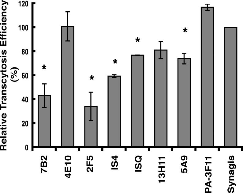
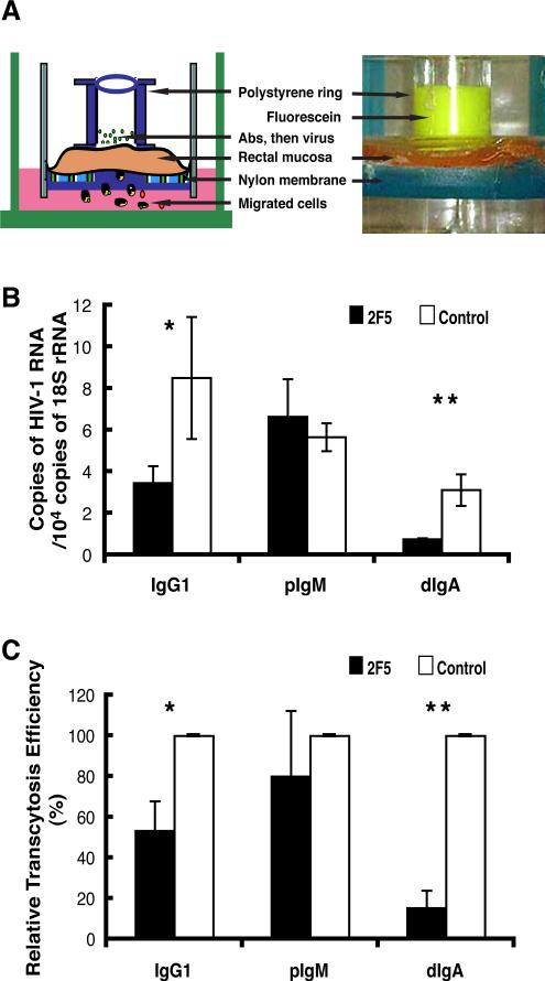
Monostratified epithelial cells translocate HIV type 1 (HIV-1) from the apical to the basolateral surface via vesicular transcytosis. Because acutely transmitted HIV-1 is almost exclusively CCR5-tropic and human intestinal epithelial cells preferentially transcytose CCR5-tropic virus, we established epithelial monolayers using polarized HT-29 cells transduced to express CCR5, and an explant system using normal human rectal mucosa, to characterize biological parameters of epithelial cell transcytosis of HIV-1 and assess antiviral Ab blockade of transcytosis. The amount of cell-free HIV-1 transcytosed through the epithelial monolayer increased linearly in relation to the amount of virus applied to the apical surface, indicating transcytosis efficiency was constant (r(2) = 0.9846; p < 0.0001). The efficiency of HIV-1 transcytosis ranged between 0.05 and 1.21%, depending on the virus strain, producer cell type and gp120 V1-V3 loop signature. Inoculation of HIV-1 neutralizing Abs to the immunodominant region (7B2) or the conserved membrane proximal external region (2F5) of gp41 or to cardiolipin (IS4) onto the apical surface of epithelial monolayers prior to inoculation of virus significantly reduced HIV-1 transcytosis. 2F5 was the most potent of these IgG1 Abs. Dimeric IgA and monomeric IgA, but not polymeric IgM, 2F5 Abs also blocked HIV-1 transcytosis across the epithelium and, importantly, across explanted normal human rectal mucosa, with monomeric IgA substantially more potent than dimeric IgA in effecting transcytosis blockade. These findings underscore the potential role of transcytosis blockade in the prevention of HIV-1 transmission across columnar epithelium such as that of the rectum.
Figures


Similar articles
-
Natural mucosal antibodies reactive with first extracellular loop of CCR5 inhibit HIV-1 transport across human epithelial cells.AIDS. 2007 Jan 2;21(1):13-22. doi: 10.1097/QAD.0b013e328011049b. AIDS. 2007. PMID: 17148963
-
Secretory IgA specific for a conserved epitope on gp41 envelope glycoprotein inhibits epithelial transcytosis of HIV-1.J Immunol. 2001 May 15;166(10):6257-65. doi: 10.4049/jimmunol.166.10.6257. J Immunol. 2001. PMID: 11342649
-
Neutralizing monoclonal antibodies to human immunodeficiency virus type 1 do not inhibit viral transcytosis through mucosal epithelial cells.Virology. 2008 Jan 20;370(2):246-54. doi: 10.1016/j.virol.2007.09.006. Epub 2007 Oct 24. Virology. 2008. PMID: 17920650
-
Infectious human immunodeficiency virus can rapidly penetrate a tight human epithelial barrier by transcytosis in a process impaired by mucosal immunoglobulins.J Infect Dis. 1999 May;179 Suppl 3:S448-53. doi: 10.1086/314802. J Infect Dis. 1999. PMID: 10099117 Review.
-
Interactions of viruses and microparticles with apical plasma membranes of M cells: implications for human immunodeficiency virus transmission.J Infect Dis. 1999 May;179 Suppl 3:S441-3. doi: 10.1086/314800. J Infect Dis. 1999. PMID: 10099115 Review.
Cited by
-
Human seminal plasma stimulates the migration of CD11c+ mononuclear phagocytes to the apical side of the colonic epithelium without altering the junctional complexes in an ex vivo human intestinal model.Front Immunol. 2023 Mar 22;14:1133886. doi: 10.3389/fimmu.2023.1133886. eCollection 2023. Front Immunol. 2023. PMID: 37033941 Free PMC article.
-
Scarcity or absence of humoral immune responses in the plasma and cervicovaginal lavage fluids of heavily HIV-1-exposed but persistently seronegative women.AIDS Res Hum Retroviruses. 2011 May;27(5):469-86. doi: 10.1089/aid.2010.0169. Epub 2010 Nov 22. AIDS Res Hum Retroviruses. 2011. PMID: 21091128 Free PMC article.
-
HIV-1 envelope glycan moieties modulate HIV-1 transmission.J Virol. 2014 Dec;88(24):14258-67. doi: 10.1128/JVI.02164-14. Epub 2014 Oct 1. J Virol. 2014. PMID: 25275130 Free PMC article.
-
Identification of Human Anti-HIV gp160 Monoclonal Antibodies That Make Effective Immunotoxins.J Virol. 2017 Jan 18;91(3):e01955-16. doi: 10.1128/JVI.01955-16. Print 2017 Feb 1. J Virol. 2017. PMID: 27852851 Free PMC article.
-
Rapid SIV Env-specific mucosal and serum antibody induction augments cellular immunity in protecting immunized, elite-controller macaques against high dose heterologous SIV challenge.Virology. 2011 Mar 1;411(1):87-102. doi: 10.1016/j.virol.2010.12.033. Epub 2011 Jan 14. Virology. 2011. PMID: 21237474 Free PMC article.
References
-
- Royce RA, Sena A, Cates W, Jr., Cohen MS. Sexual transmission of HIV. N. Engl. J. Med. 1997;336:1072–1078. - PubMed
-
- Wawer MJ, Gray RH, Sewankambo NK, Serwadda D, Li X, Laeyendecker O, Kiwanuka N, Kigozi G, Kiddugavu M, Lutalo T, Nalugoda F, Wabwire-Mangen F, Meehan MP, Quinn TC. Rates of HIV-1 transmission per coital act, by stage of HIV-1 infection, in Rakai, Uganda. J Infect Dis. 2005;191:1403–1409. - PubMed
-
- Shattock RJ, Moore JP. Inhibiting sexual transmission of HIV-1 infection. Nat. Rev. Microbiol. 2003;1:25–34. - PubMed
Publication types
MeSH terms
Substances
Grants and funding
- U01 AI067854/AI/NIAID NIH HHS/United States
- R21 AI083539/AI/NIAID NIH HHS/United States
- DK-47322/DK/NIDDK NIH HHS/United States
- R24 DK064400/DK/NIDDK NIH HHS/United States
- RR-20136/RR/NCRR NIH HHS/United States
- C06 RR020136/RR/NCRR NIH HHS/United States
- AI-83027/AI/NIAID NIH HHS/United States
- R01 DK054495/DK/NIDDK NIH HHS/United States
- AI-83539/AI/NIAID NIH HHS/United States
- AI-67854/AI/NIAID NIH HHS/United States
- U19 AI067854/AI/NIAID NIH HHS/United States
- R01 DK047322/DK/NIDDK NIH HHS/United States
- DK-64400/DK/NIDDK NIH HHS/United States
- P01 AI083027/AI/NIAID NIH HHS/United States
- I01 CX000372/CX/CSRD VA/United States
- DK-54495/DK/NIDDK NIH HHS/United States
LinkOut - more resources
Full Text Sources
Other Literature Sources
Medical
Miscellaneous
