. 2010 Apr 1;184(7):4033-41.
doi: 10.4049/jimmunol.0903069. Epub 2010 Mar 5.

Coordinate interaction between IL-13 and epithelial differentiation cluster genes in eosinophilic esophagitis

Affiliations

Coordinate interaction between IL-13 and epithelial differentiation cluster genes in eosinophilic esophagitis

Carine Blanchard et al. J Immunol. .

Abstract

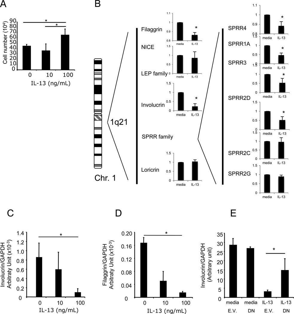
We have previously proposed that the pathogenesis of eosinophilic esophagitis (EE) is mediated by an IL-13-driven epithelial cell response associated with marked gene dysregulation including eotaxin-3 overproduction. In this study, we compared epithelial responses between healthy patients and those with EE, aiming to uncover molecular explanations for EE pathogenesis. Esophageal epithelial cells could be maintained for up to five passages, with 67% and 62% of cell lines reaching confluence in healthy controls and EE cases, respectively. Both sets of epithelial cells avidly responded to IL-13 at similar levels as assessed by eotaxin-3 production. Acidic pH increased cellular release of eotaxin-3 (4.6 +/- 1.98 ng/ml versus 12.46 +/- 2.90 ng/ml at pH 7.4 and 4, respectively; p < 0.05). Numerous epidermal differentiation complex (EDC) genes, such as filaggrin and SPRR3, were downregulated both in IL-13-stimulated esophageal epithelial cells and in EE biopsies specimens compared with healthy controls. Whereas the filaggrin loss of function mutation 2282del4 was overrepresented in EE compared with control individuals (6.1% versus 1.3% respectively; p = 0.0172), the decreased filaggrin expression was uniformly seen in all EE cases in vivo. Indeed, expression of the EDC genes filaggrin and involucrin was strongly decreased directly by IL-13. These results establish that the epithelial response in EE involves a cooperative interaction between IL-13 and expression of EDC genes.


Figures

Figure 1
Figure 1. Growth characteristics and response to IL-13 of primary esophageal epithelial cells from normal (NL) and eosinophilic esophagitis (EE) patients
Cells were grown in modified F-media for a maximum of 35 days. In A, the percentage of cells that reach 80-100% confluence is shown in the NL and EE group. In B, the number of cells at confluence or after 35 days of culture is presented. In C, the number of days to reach 80-100% confluence is shown. In D, the fold increase in eotaxin-3 mRNA expression following IL-13 stimulation at different doses is shown; note the Log10 scale. In D, data points are average ± SD, non significant (ns). In E, EE and NL derived cells were stimulated for 48h with IL-13 (100 ng/mL) then the media was acidified or heparinized for 4h and eotaxin-3 protein was quantified in the supernatant. Bars indicate mean values ± SD, * p<0.05.
Figure 2
Figure 2. Chromosome involvement in the EE transcriptome and physical map of the epidermal differentiation complex gene cluster on the chromosome 1
In A, the EE transcriptome was assigned to chromosomes using DAVID ontology. The number of genes and the p value are presented. In B, a schematics of the physical map of the 1q21 gene cluster and the corresponding microarray expression profile in NL and EE patients are shown.
Figure 3
Figure 3. Esophageal expression of filaggrin mRNA
A. Filaggrin expression was assessed by real-time PCR and normalized to GAPDH in control patients (NL), active EE patients (EE active; eosinophil level ≥ 24), EE patients with an intermediate eosinophilia level (EE int; eosinophils level from 1 to 23) and inactive EE patients who responded successfully to their therapy (inactive; eosinophil level = 0). N = 48, 21, 18 2 and 9 individuals in EE active, EE intermediate, EE inactive and NL groups, respectively. Each data point corresponds to a patient and the lines show the median with interquartile range. C and D Involucrin and SPRR3 expression were assessed by realtime PCR and normalized to GAPDH as described in (A). Each data point corresponds to a patient and the lines show the median with interquartile range.
Figure 4
Figure 4. Filaggrin expression in active EE patients with or without allergic symptoms or sensitization
Filaggrin expression was assessed by real-time PCR and normalized to GAPDH in non-allergic (n=7) and allergic EE (n=25) patients. Each data point corresponds to a patient and the lines show the median with the interquartile range.
Figure 5
Figure 5. Physical map and gene expression of the epidermal differentiation complex gene cluster on the chromosome 1 in IL-13-stimulated primary esophageal epithelial cells
In A, the effect of chronic exposure of IL-13 on esophageal epithelial cell number is shown. Cells (10% confluence) were stimulated every 3 days with IL-13 (0, 10, 100 ng/mL) for 9 days (80-90% confluence). Cell numbers are presented as average ± SD. The results are representative of at least 3 independent experiments. In B, schematics of the physical map of the 1q21 gene cluster and the microarray expression profile primary esophageal epithelial cells stimulated with IL-13. In B and C, esophageal primary epithelial cells from EE patients were stimulated with IL-13 (100 ng/mL) for 48h. Total mRNA was extracted and subjected to real-time RT-PCR for involucrin (B) and filaggrin (C). In D, the esophageal epithelial cell line TE-7 (<10% confluence), was transfected with a dominant negative form of STAT6 (DN) or the empty vector (E.V.). Total mRNA was extracted and submitted to real-time RT-PCR for involucrin mRNA quantification. Results are normalized to a housekeeping gene (GAPDH). Bars are average ± SD. Experiments were performed in triplicate, * is p<0.05.

References

    1. Furuta GT, Liacouras CA, Collins MH, Gupta SK, Justinich C, Putnam PE, Bonis P, Hassall E, Straumann A, Rothenberg ME. Eosinophilic esophagitis in children and adults: a systematic review and consensus recommendations for diagnosis and treatment. Gastroenterology. 2007;133:1342–1363. - PubMed
    1. Molina-Infante J, Ferrando-Lamana L, Mateos-Rodriguez JM, Perez-Gallardo B, Prieto-Bermejo AB. Overlap of reflux and eosinophilic esophagitis in two patients requiring different therapies: a review of the literature. World J Gastroenterol. 2008;14:1463–1466. - PMC - PubMed
    1. Lierse W. The physiology and pathology of the esophagus. Eur J Pediatr Surg. 1992;2:323–326. - PubMed
    1. De Benedetto A, Qualia CM, Baroody FM, Beck LA. Filaggrin expression in oral, nasal, and esophageal mucosa. J Invest Dermatol. 2008;128:1594–1597. - PubMed
    1. Crish JF, Bone F, Banks EB, Eckert RL. The human involucrin gene contains spatially distinct regulatory elements that regulate expression during early versus late epidermal differentiation. Oncogene. 2002;21:738–747. - PubMed

Publication types

MeSH terms