. 2010 May 14;285(20):15065-15075.
doi: 10.1074/jbc.M109.087445. Epub 2010 Mar 5.

Cell surface relocalization of the endoplasmic reticulum chaperone and unfolded protein response regulator GRP78/BiP

Affiliations

Cell surface relocalization of the endoplasmic reticulum chaperone and unfolded protein response regulator GRP78/BiP

Yi Zhang et al. J Biol Chem. .

Abstract

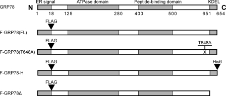
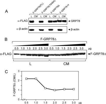
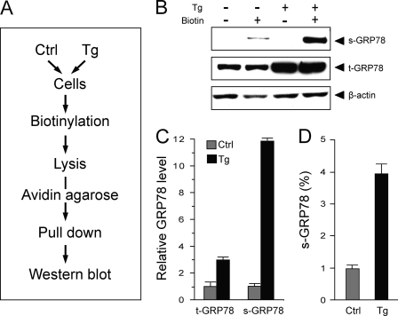
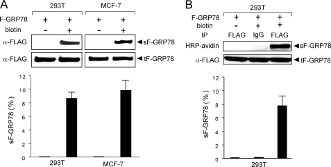
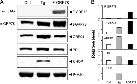
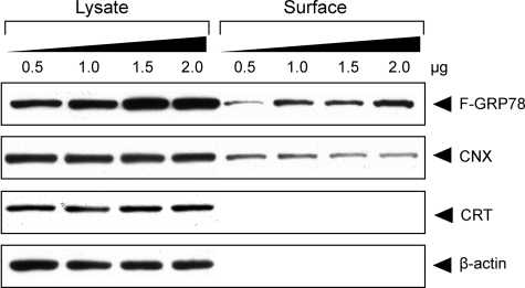
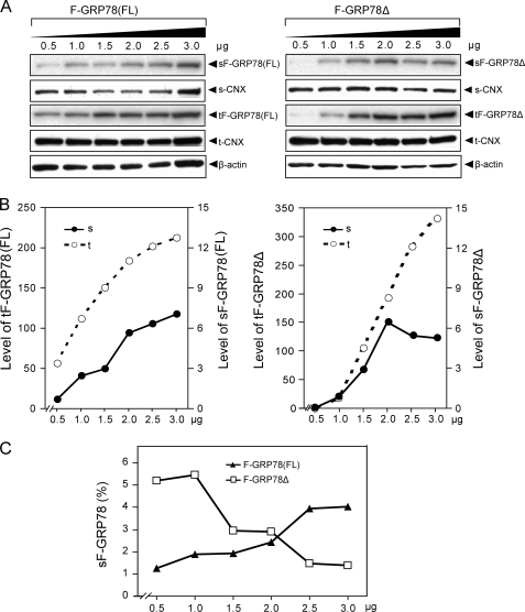
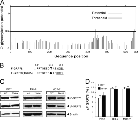
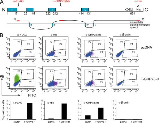
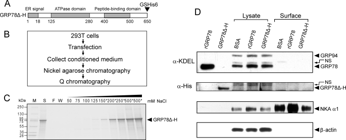
The recent discovery that GRP78/BiP, a typical endoplasmic reticulum (ER) lumenal chaperone, can be expressed on the cell surface, interacting with an increasing repertoire of surface proteins and acting as receptor in signaling pathways, represents a paradigm shift in its biological function. However, the mechanism of GRP78 trafficking from the ER to the cell surface is not well understood. Using a combination of cellular, biochemical, and mutational approaches, we tested multiple hypotheses. Here we report that ER stress actively promotes GRP78 localization on the cell surface, whereas ectopic expression of GRP78 is also able to cause cell surface relocation in the absence of ER stress. Moreover, deletion of the C-terminal ER retention motif in GRP78 alters its cell surface presentation in a dose-dependent manner; however, mutation of the putative O-linked glycosylation site Thr(648) of human GRP78 is without effect. We also identified the exposure of multiple domains of GRP78 on the cell surface and determined that binding of extracellular GRP78 to the cell surface is unlikely. A new topology model for cell surface GRP78 is presented.


Figures

FIGURE 1.
FIGURE 1.
Schematic illustration of the wild type and mutated forms of human GRP78 encoded by the expression plasmids. The ER signal peptide, ATPase domain, peptide-binding domain, and KDEL motif are indicated. The FLAG sequence was inserted immediately after the ER signal peptide. The full-length, wild type protein was denoted as F-GRP78(FL). For glycosylation site mutant F-GRP78(T648A), threonine at position 648 was exchanged to alanine. For F-GRP78-H, six tandem histidine (His6) was added following the KDEL motif of F-GRP78(FL). For F-GRP78Δ, the KDEL motif was deleted from F-GRP78(FL).
FIGURE 2.
FIGURE 2.
Measurement of intracellular and secreted GRP78 in wild type and mutant proteins. A, 293T cells were transfected with F-GRP78(FL), F-GRP78(T648A), F-GRP78Δ, or F-GRP78-H. After 6 h, the cells were grown in hybridoma serum-free medium and collected after 48 h. 5% total cell lysate (L) and concentrated conditioned medium (CM) were subjected to Western blot for detection of FLAG-tagged GRP78. β-Actin served as a loading control for the lysates and a test for cell lysis resulting in leakage of intracellular protein into conditioned medium. B, 293T cells were transfected with increasing amounts of F-GRP78Δ DNA in 6-cm dishes. pcDNA was added to equalize the amount of DNA for transfection. 5% total cell lysate and concentrated conditioned medium were subjected to Western blot for detection of F-GRP78Δ. C, after quantitation of B, the ratio of secreted F-GRP78Δ in conditioned medium versus total intracellular F-GRP78Δ in lysate was plotted against the dosage of expression plasmid.
FIGURE 3.
FIGURE 3.
ER stress actively promotes cell surface localization of endogenous GRP78. A, flow chart of the strategy to detect cell surface protein in cells that are either untreated (Ctrl) or subjected to Tg treatment. B, detection of cell surface GRP78 expression in 293T cells either untreated (−) or treated (+) with 300 nm Tg for 16 h. β-Actin served as loading control. Representative Western blots are shown. s-GRP78 and t-GRP78, surface and total intracellular GRP78, respectively. The amount of total lysate was 10% of the amount used for avidin pull-down. C, quantitation of the relative expression level of total intracellular GRP78 and cell surface GRP78 in control and Tg-treated cells. D, the fraction of cell surface versus total intracellular GRP78 in control and Tg-treated cells. The experiments were repeated 3–4 times. The S.D. is shown.
FIGURE 4.
FIGURE 4.
Detection of cell surface GRP78 following ectopic expression in 293T and MCF-7 cells. The cells were transfected with 2.0 μg of F-GRP78 in 6-well dishes, and either biotinylated or non-biotinylated. The cell lysates were subjected to avidin pull-down (A) or immunoprecipitation (IP) with anti-FLAG antibody (B), followed by Western blots with anti-FLAG antibody or developed with horseradish peroxidase-avidin, as indicated. The amount of total lysate was 10% of the amount used for avidin pull-down or immunoprecipitation. Representative Western blots are shown. sF-GRP78 and tF-GRP78, surface and total intracellular F-GRP78, respectively. After quantitation, the percentage of cell surface F-GRP78 was calculated, and it is presented below the autoradiogram. The experiments were repeated 3–4 times. The S.D. is shown.
FIGURE 5.
FIGURE 5.
Ectopic expression of GRP78 does not induce ER stress. A, 293T cells were either treated with Tg (300 nm) for 16 h or transfected with full-length FLAG-tagged GRP78 for 48 h. Cells without Tg treatment and transfection served as control (Ctrl). Total cell lysate was prepared and subjected to Western blot analysis. The protein levels of exogenous GRP78 (F-GRP78); total intracellular GRP78 (t-GRP78); endogenous GRP94, PDI, and CHOP; and β-actin, which served as loading control, are shown. B, quantitation of the protein band intensities shown in A, with the level of the control cells set as 1.
FIGURE 6.
FIGURE 6.
Dosage-dependent increase of surface expression of GRP78. 293T cells were transfected with the indicated amount of F-GRP78 expression plasmid. F-GRP78 and CNX were detected on the cell surface by Western blot, whereas calreticulin (CRT) was barely detected on the cell surface. β-Actin served as loading control for the input lysate, which represents 10% of the amount used for avidin pull-down to detect surface protein.
FIGURE 7.
FIGURE 7.
Deletion of the ER retrieval signal, KDEL, affects cell surface localization of GRP78. A, 293T cells in 6-cm dishes were transfected with increasing amounts of F-GRP78(FL) or F-GRP78Δ, as indicated, and pcDNA vector was added to equalize total DNA amount in each transfection. Cell surface proteins were purified and analyzed as described in the legend to Fig. 6. s, surface proteins; t, lysate input. B, the relative level of F-GRP78(FL) and F-GRP78Δ on cell surface or in total lysate. After quantitation of protein band intensity, surface F-GRP78(FL) and surface F-GRP78-Δ were normalized against surface CNX, whereas total intracellular F-GRP78(FL) and F-GRP78-Δ were normalized against β-actin. The levels of surface and total intracellular F-GRP78(FL) or F-GRP78Δ are plotted against the dosage of the expression plasmid. C, the percentages of cell surface F-GRP78(FL) and F-GRP78Δ were calculated and plotted against the dosage of the expression plasmid.
FIGURE 8.
FIGURE 8.
Mutation of O-linked glycosylation site (T648A) does not affect cell surface translocation of GRP78. A, schematic diagram of O-linked glycosylation sites predicted by the Net OGly 3.1 program for human GRP78. The threshold line of glycosylation potential is indicated as a black boldface line. The amino acid sequence position is shown below. B, the amino acid sequences surrounding the putative O-linked glycosylation site at aa 648 of human GRP78, with the mutated site indicated in boldface type. C, detection of surface and total intracellular F-GRP78 (wild type and mutant) in MCF7, HeLa, and 293T cells. β-Actin served as the loading control for the lysate input. Representative Western blots are shown. D, after quantitation of protein band intensity, the percentages of cell surface F-GRP78 (sF-GRP78; wild type (WT) and mutant) in the different cell lines were calculated and are presented. The experiments were repeated 3–4 times. The S.D. is shown.
FIGURE 9.
FIGURE 9.
Multiple domains of GRP78 are exposed on cell surface. HeLa cells transfected with F-GRP78-H containing N-terminal FLAG tag and C-terminal His tag were subjected to FACS analysis. Cells transfected with pcDNA vector were used as negative control. A, schematic drawing of human GRP78 with the potential transmembrane domains (I–IV) predicted by TMPred (ExPasy tools) indicated in blue. The ER signal sequence is indicated as an asterisk. The numbers below refer to the amino acid residues. The domains recognized by the antibodies used for FACS analysis are indicated in red. The α-GRP78 (M) antibody recognizes the middle domain of human GRP78 spanning aa 250–300. Below is a proposed topology model of cell surface GRP78, with the putative transmembrane domains indicated in blue and the extracellular domains identified by FACS in red. B, exposure of the N terminus, C terminus, and middle domain (aa 250–300) of GRP78 at the cell surface was detected by FACS analysis using the antibodies as indicated. β-Actin was used as control for cell integrity. The percentage of positive cells is shown under each FACS profile. PE, phycoerythrin; FITC, fluorescein isothiocyanate.
FIGURE 10.
FIGURE 10.
Extracellular GRP78 does not stably bind to cell surface. A, schematic illustration of GRP78Δ-H mutant encoded by expression plasmid, in which KDEL motif was deleted and glycine (G) and serine (S), followed by six tandem histidine (GSHis6), were added at the C terminus of GRP78. B, methodological flow chart of expression and purification of secreted GRP78Δ-H. C, Q chromatography purification for GRP78Δ-H. Protein purified by nickel-agarose affinity chromatography was loaded onto a Q column, and after extensive wash of the column, elution was performed with buffer containing the indicated concentration (50–500 mm) of NaCl. All fractions were run on SDS-PAGE followed by Coomassie Blue staining. The fractions marked with an asterisk were collected and saved. The molecular weight of protein marker (M) is indicated on the left. S, protein initially loaded onto the Q column; F, flow-through fraction; W, wash fraction. D, detection of extracellular GRP78 entering or binding to surface of 293T cells. 10 μg of rGRP78, purified GRP78Δ-H, or bovine serum albumin (BSA) were added in the medium of 293T cells in 6-well dishes, respectively, and incubated for 24 h. The cell surface and 20% lysate input of GRP78 and GRP78Δ-H were detected by Western blot against either anti-KDEL or anti-His antibody. 1 μg of rGRP78 and purified GRP78Δ-H were loaded as positive control. The upper band above the positive control is a nonspecific (NS) band. Sodium-potassium-ATPase (NKA α1) served as cell surface protein loading control. β-Actin was used as lysate input loading control.

Similar articles

Cited by

References

    1. Ni M., Lee A. S. (2007) FEBS Lett. 581, 3641–3651 - PMC - PubMed
    1. Hendershot L. M. (2004) Mt. Sinai J. Med. 71, 289–297 - PubMed
    1. Lee A. S. (2005) Methods 35, 373–381 - PubMed
    1. Fu Y., Lee A. S. (2006) Cancer Biol. Ther. 5, 741–744 - PubMed
    1. Lee A. S. (2007) Cancer Res. 67, 3496–3499 - PubMed

Publication types