. 2010 Mar 1;66(Pt 3):242-50.
doi: 10.1107/S1744309109055158. Epub 2010 Feb 23.

Apo and ligand-bound structures of ModA from the archaeon Methanosarcina acetivorans

Affiliations

Apo and ligand-bound structures of ModA from the archaeon Methanosarcina acetivorans

Sum Chan et al. Acta Crystallogr Sect F Struct Biol Cryst Commun. .

Abstract

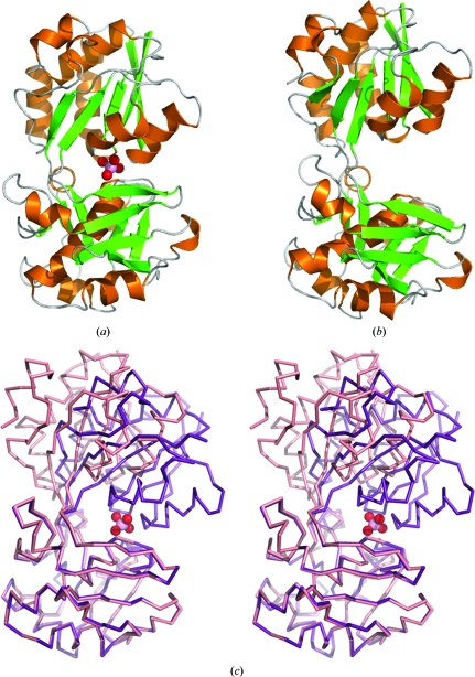
The trace-element oxyanion molybdate, which is required for the growth of many bacterial and archaeal species, is transported into the cell by an ATP-binding cassette (ABC) transporter superfamily uptake system called ModABC. ModABC consists of the ModA periplasmic solute-binding protein, the integral membrane-transport protein ModB and the ATP-binding and hydrolysis cassette protein ModC. In this study, X-ray crystal structures of ModA from the archaeon Methanosarcina acetivorans (MaModA) have been determined in the apoprotein conformation at 1.95 and 1.69 A resolution and in the molybdate-bound conformation at 2.25 and 2.45 A resolution. The overall domain structure of MaModA is similar to other ModA proteins in that it has a bilobal structure in which two mixed alpha/beta domains are linked by a hinge region. The apo MaModA is the first unliganded archaeal ModA structure to be determined: it exhibits a deep cleft between the two domains and confirms that upon binding ligand one domain is rotated towards the other by a hinge-bending motion, which is consistent with the 'Venus flytrap' model seen for bacterial-type periplasmic binding proteins. In contrast to the bacterial ModA structures, which have tetrahedral coordination of their metal substrates, molybdate-bound MaModA employs octahedral coordination of its substrate like other archaeal ModA proteins.


Figures

Figure 1
Figure 1
Comparison of apo and molybdate-bound MaModA. (a) Ribbon diagram of the molybdate-bound conformation of MaModA (PDB code 3k6w). β-­Strands are shown in green, α-helices in orange and loops in gray. The molybdate ligand is shown in space-filling representation with the O atoms colored red and the molybdenum in pink. (b) Ribbon diagram of the apo conformation of MaModA (PDB code 3k6u) colored as in (a). (c) Superposition of the ligand-bound and apo conformations of the MaModA backbone shown in stereo stick representation. The ligand-bound conformation is colored purple and the apo conformation is in salmon. The molybdate is colored as in (a).
Figure 2
Figure 2
The β-barrel formed by the four-stranded β-sheet appendage. Two views are shown of the two ligand-bound MaModA molecules found in the asymmetric unit of crystal form II. The β-strands that contribute to the β-barrel are shown in ribbon representation, while the remaining main-chain residues are shown in stick representation.
Figure 3
Figure 3
Superposition stereoview of the ligand-binding sites of the molybdate-bound MaModA structure (PDB code 3k6x) and the tungstate-bound MaModA structure (PDB code 3cfx). (a) Molybdate-bound MaModA is shown in ribbon representation and is colored gray, with the molybdenum colored cyan. The tungstate-bound MaModA is shown in pale green as is the tungstate ligand. Residues that interact with the ligands are shown in stick representation and bonds between the molybdate-bound structure and its ligand are shown by dashed lines. (b) Two-dimensional representation of the octahedral coordination of the molybdate substrate by MaModA.
Figure 4
Figure 4
Sequence alignment of MaModA, E. coli ModA and archaeal ModA homologues for which structures have been determined. Two additional proteins from M. acetivorans (MA1237 and MA2280) that have been annotated as ModA proteins but whose structures have not yet been determined are included. Secondary-structure elements for molybdate-bound MaModA (MA0280, PDB codes 3k6w and 3k6x, this study) are shown above the alignment. Amino-acid residues that have been determined to interact with either molybdate or tungstate ligands from cocrystal structures are indicated as follows: blue, interaction with ligand via backbone amine; red, interaction with ligand via side chain; green, interaction with ligand via both backbone amine and side chain. Identical residues are shaded in dark gray and conserved residues are shaded in light gray. Protein sequences are MA0280, Methanosarcina acetivorans ModA; Archaeoglobus fulgidus ModA, PDB code 2onr; Methanocaldococcus jannaschii ModA, PDB code 3cfz; Pyrococcus horikoshii ModA, PDB code 3cg3; P. furiosus ModA, PDB code 3cg1; E. coli ModA, PDB code 1amf.

Similar articles

Cited by

References

    1. Austermuhle, M. I., Hall, J. A., Klug, C. S. & Davidson, A. L. (2004). J. Biol. Chem.279, 28243–28250. - PubMed
    1. Balan, A., Santacruz-Perez, C., Moutran, A., Ferreira, L. C., Neshich, G. & Barbosa, J. A. (2008). Biochim. Biophys. Acta, 1784, 393–399. - PubMed
    1. Brylinski, M. & Skolnick, J. (2008). Proteins, 70, 363–377. - PubMed
    1. Clarke, T. E., Ku, S. Y., Dougan, D. R., Vogel, H. J. & Tari, L. W. (2000). Nature Struct. Biol.7, 287–291. - PubMed
    1. Collaborative Computational Project, Number 4 (1994). Acta Cryst. D50, 760–763. - PubMed

Publication types

MeSH terms