Comparative Study
. 2010 Apr;13(4):507-12.
doi: 10.1038/nn.2509. Epub 2010 Mar 7.

A central role for the lateral prefrontal cortex in goal-directed and stimulus-driven attention

Affiliations
Comparative Study

A central role for the lateral prefrontal cortex in goal-directed and stimulus-driven attention

Christopher L Asplund et al. Nat Neurosci. 2010 Apr.

Abstract

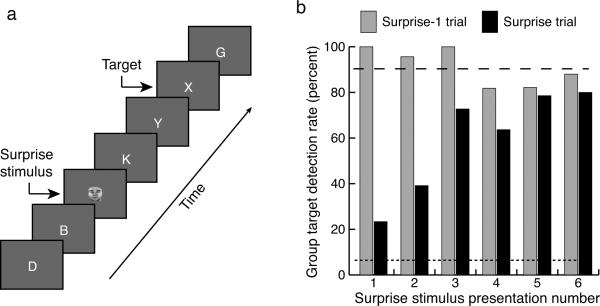
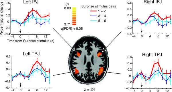
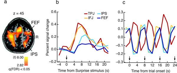
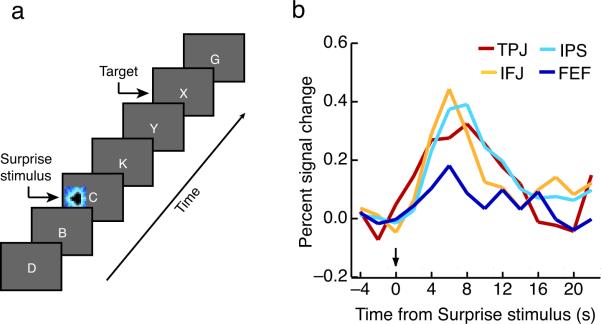
Attention is the process that selects which sensory information is preferentially processed and ultimately reaches our awareness. Attention, however, is not a unitary process; it can be captured by unexpected or salient events (stimulus driven) or it can be deployed under voluntary control (goal directed), and these two forms of attention are implemented by largely distinct ventral and dorsal parieto-frontal networks. For coherent behavior and awareness to emerge, stimulus-driven and goal-directed behavior must ultimately interact. We found that the ventral, but not dorsal, network can account for stimulus-driven attentional limits to conscious perception, and that stimulus-driven and goal-directed attention converge in the lateral prefrontal component of that network. Although these results do not rule out dorsal network involvement in awareness when goal-directed task demands are present, they point to a general role for the lateral prefrontal cortex in the control of attention and awareness.


Figures

Figure 1
Figure 1
SiB experiment (Experiment 1). A) Trial design. Participants searched for a target letter in a rapid serial visual presentation (RSVP) stream of distractor letters. In a small proportion of trials (Surprise trials), a Surprise face stimulus was shown before the target. B) Group target detection performance. Black bars represent accuracy in Surprise trials, and gray bars represent accuracy in trials immediately preceding the Surprise trials. Dashed line corresponds to the average target hit rate for Search trials (target only). Dotted line corresponds to the false alarm rate.
Figure 2
Figure 2
SiB experiment (Experiment 1) SPM. Brain regions showing rapid attenuation of Surprise stimulus-related activation. The SPM highlights brain regions that responded to all six Surprise trials (See Methods), specifically the IFJs (Talairach coordinates 37, 5, 29 and −40, 8, 25) and TPJs (Talairach coordinates 46, −56, 27 and −49, −56, 23). The time courses illustrate the brain regions from the SPM that showed greater activity in the first pair of Surprise trials compared to the two other pairs of Surprise trials. The Surprise stimulus appears at approximately time zero. Error bars represent standard errors of the mean.
Figure 3
Figure 3
Stimulus-driven and goal-directed attention activity in Experiment 1. A) Dorsal brain regions active during Search trials. B) Surprise stimulus-specific waveform in dorsal (FEF, IPS) and ventral (IFJ, TPJ) regions of interest (ROIs) defined in individual participants (See Methods). Each time course was constructed by subtracting the Search trial time course from the time course for the first two Surprise Stimulus trials. The Surprise stimulus appears at approximately time zero. C) Search trial time course over the same period of time for the same ROIs. Arrows mark each trial's onset. Note that the activation pattern is cyclical, mirroring the trial structure (one trial every eight seconds). The observed hemodynamic responses match the predicted responses for the hypothesized neural activity in each region (see Supplementary Fig. 3).
Figure 4
Figure 4
Spatial SiB Experiment (Experiment 2). A) Trial design. The procedure was identical to that in Experiment 1 save that in a small proportion of trials, a colorful Surprise stimulus was shown before the target away from fixation (see Fig. 1a). B) Surprise stimulus-specific waveforms in dorsal and ventral attention network ROIs defined in individual participants. Time courses were constructed in the same fashion as those in Experiment 1 (see Fig. 3b, main text). Note that all four regions show an immediate response to the Surprise stimulus presentations.
Figure 5
Figure 5
Endogenous Cueing Task Experiment (Experiment 3). A) Trial design. A color cue predicted the location of an upcoming target, to which the participant then responded in a speeded manner. B) Cue-related activity in dorsal and ventral attention network ROIs isolated from Experiment 2 (See Methods). The arrow marks cue onset.

Similar articles

Cited by

References

    1. Corbetta M, Shulman GL. Control of goal-directed and stimulus-driven attention in the brain. Nature Reviews Neuroscience. 2002;3:201–215. - PubMed
    1. Egeth HE, Yantis S. Visual attention: control, representation, and time course. Annual Review of Psychology. 1997;48:269–297. - PubMed
    1. Corbetta M, Kincade JM, Ollinger JM, McAvoy MP, Shulman GL. Voluntary orienting is dissociated from target detection in human posterior parietal cortex. Nature Neuroscience. 2000;3:292–297. - PubMed
    1. Kastner S, Pinsk MA, De Weerd P, Desimone R, Ungerleider LG. Increased activity in human visual cortex during directed attention in the absence of visual stimulation. Neuron. 1999;22:751–761. - PubMed
    1. Serences JT, Shomstein S, Leber AB, Golay X, et al. Coordination of voluntary and stimulus-driven attentional control in human cortex. Psychological Science. 2005;16:114–122. - PubMed

Publication types