. 2010 Apr;16(4):446-51.
doi: 10.1038/nm.2109. Epub 2010 Mar 7.

HIV-1 infects multipotent progenitor cells causing cell death and establishing latent cellular reservoirs

Affiliations

HIV-1 infects multipotent progenitor cells causing cell death and establishing latent cellular reservoirs

Christoph C Carter et al. Nat Med. 2010 Apr.

Abstract

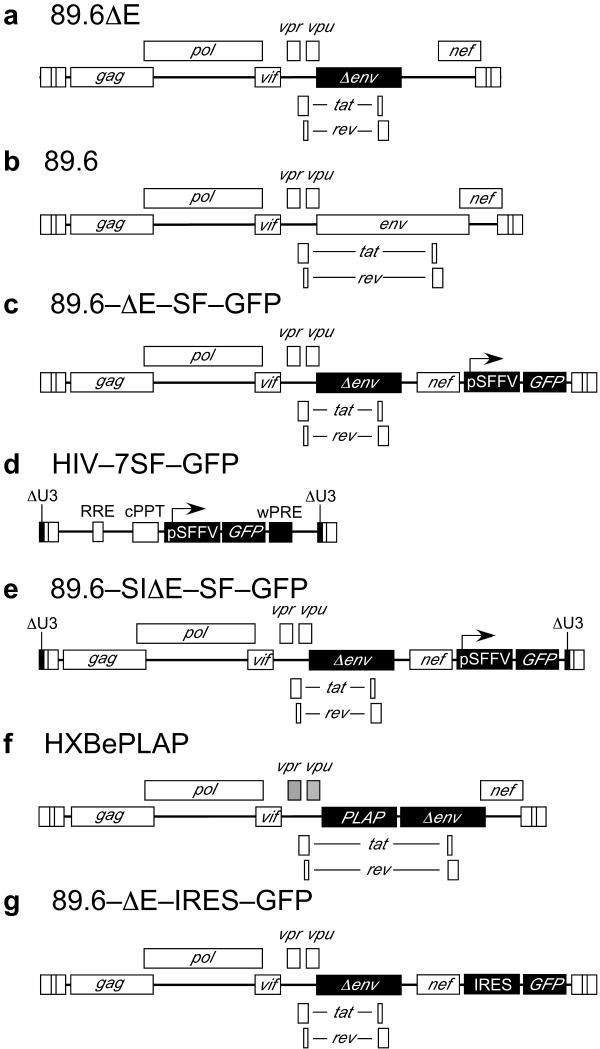
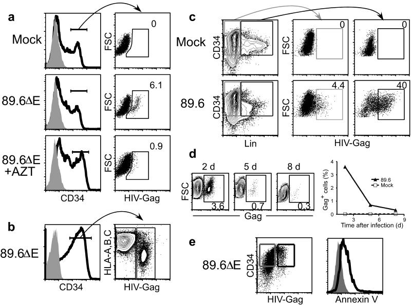
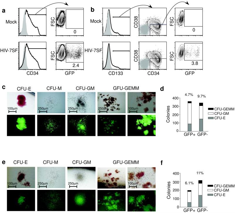
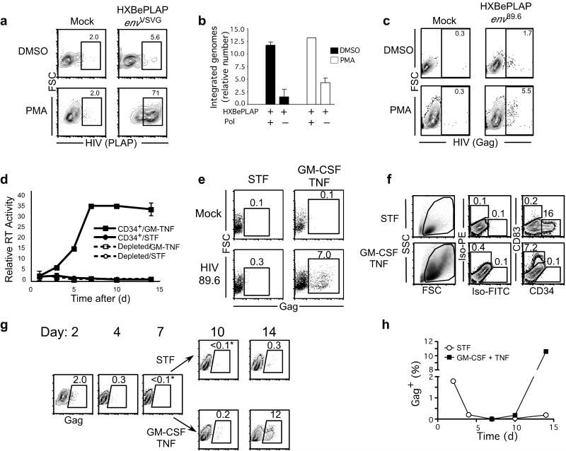
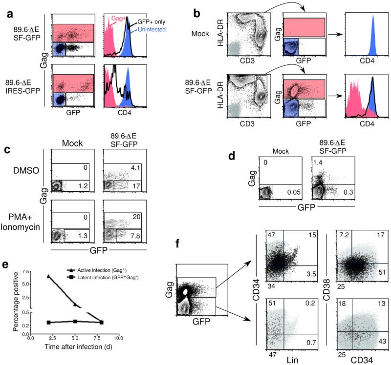
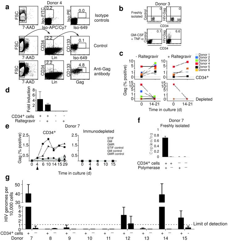
HIV causes a chronic infection characterized by depletion of CD4(+) T lymphocytes and the development of opportunistic infections. Despite drugs that inhibit viral spread, HIV infection has been difficult to cure because of uncharacterized reservoirs of infected cells that are resistant to highly active antiretroviral therapy (HAART) and the immune response. Here we used CD34(+) cells from infected people as well as in vitro studies of wild-type HIV to show infection and killing of CD34(+) multipotent hematopoietic progenitor cells (HPCs). In some HPCs, we detected latent infection that stably persisted in cell culture until viral gene expression was activated by differentiation factors. A unique reporter HIV that directly detects latently infected cells in vitro confirmed the presence of distinct populations of active and latently infected HPCs. These findings have major implications for understanding HIV bone marrow pathology and the mechanisms by which HIV causes persistent infection.


Figures

Figure 1
Figure 1
HIV genomes. (a), (b), (c), (e) and (g) are derived from the molecular clone p89.6. (d) and (f) have been described elsewhere and are derived from HXB and NL4–3 -. Expressed viral genes are shown in white, deletions and additions to the genome are shown in black, and non–functional genes are shaded in gray.
Figure 2
Figure 2
HIV actively infects HPCs, leading to cell death. (a). Intracellular Gag in BM–derived CD34+ HPCs infected with HIV 89.6ΔEenv89.6 (Fig. 1a) for 3 d. Gray histograms are isotype controls. (b) CD34, MHC-I and intracellular Gag expression in UCB–derived CD34+ HPCs infected with 89.6ΔEenv89.6 for 48 h. (c) Gag, CD34 and Lin staining in BM–derived CD34+ HPCs infected with 89.6. (d) Time course of intracellular Gag expression in UCB–derived CD34+ HPCs infected with 89.6. (e) Annexin V reactivity in UCB–derived CD34+ HPCs infected with 89.6ΔEenv89.6 for 48 h. The right panel shows CD34+Gag (gray gate and histograms) and CD34+Gag+ cells (black gate and histogram).
Figure 3
Figure 3
HIV infects multipotent HPCs. (a) GFP expression in CD34+ UCB cells infected with HIV–7SF–GFPenv89.6 for 3 d. (b) The percentage of CD133+,CD34+,CD38 cells expressing GFP 3 d post–infection with HIV–7SF–GFPenv89.6. Gray histograms and events represent isotype control staining. (c) and (e), Colony formation by GFP+CD133+ UCB–derived HPCs infected with HIV–7SF–GFPenv89.6(c) or 89.6–SIΔE–GFPenv89.6 (e). (d) and (f), The relative number of colonies formed by equal numbers of sorted GFP+ and GFP, CD133+ UCBs infected with HIV–7SF–GFPenv89.6 (d) or 89.6–SIΔE–GFPenv89 (f). (erythroid (CFU–E), granulocyte–macrophage (CFU–GM), multi–lineage (CFU–GEMM)).
Figure 4
Figure 4
Induction of HIV from latency. (a) The percentage of BM CD34+ HPCs (See Supplementary Fig. 2b for initial cell purity) expressing an HIV marker gene (PLAP) following infection with HXB–ePLAPenvVSVG plus or minus PMA. (b) qPCR of integrated DNA from BM CD34+ cells infected and cultured as in (a). Pol–minus samples lack polymerase in the first round. Data are displayed as mean relative amount of integrated HIV DNA ± standard deviation (sd), n = 3. (c) The percentage of BM–derived CD34+ HPCs (Supplementary Fig. 2c) expressing intracellular Gag following infection with HXB–ePLAPenv89.6 plus or minus 10 ng ml−1 PMA. (d) Reverse transcriptase activity of CD34+ BM HPCs (Supplementary Fig. 2c) infected with HIV 89.6, plus or minus GM–CSF–TNF–α. The mean ± sd, n = 3 is shown. Control is BMMC immunodepleted for CD34. (e) Intracellular Gag expression 14 d post–infection for BM–derived HPCs infected and cultured as in (d). (f) CD34 and CD83 expression (right panels) after 2 weeks in CC110 or GM–CSF–TNF–α. Isotype–matched controls are shown (Iso–FITC and Iso–PE). (g) Intracellular Gag expression for BM–derived HPCs (Supplementary Fig. 2e) infected with HIV 89.6 and cultured in CC110 cocktail. On day 7, the cells were divided either into CC110 cocktail or GM–CSF–TNF–α. Asterisks indicate Gag reactivity < mock treated cells. (h) Graphical representation of the experiment depicted in part (g).
Figure 5
Figure 5
Active and latent infection in T cells and HPCs. (a) Gag, GFP and CD4 expression 7 d after infection in CEM–SS cells infected with 89.6–ΔE–SF–GFPenv89.6 or 89.6–ΔE–IRES–GFPenv89.6. Histogram shading corresponds to cell gate. (b) Flow cytometric analysis of PHA–activated PBMC infected with 89.6–ΔE–SF–GFPenv89.6 for 48 h. The histogram is shaded to match the gated cells. In the left panel, the isotype control is shown in gray. (c) Flow cytometric analysis of Jurkat cells infected with 89.6–ΔE–SF–GFPenv89.6 for 7 d, then split into PMA and ionomycin or DMSO control for 48 h. (d) Flow cytometric analysis of UCB–derived CD34+ HPCs infected with 89.6–ΔE–SF–GFPenv89.6 for 3 d. (e) Time course analysis of Gag+ and Gag GFP+ UCB–derived CD34+ HPCs infected as above and cultured in CC110. (f) Flow cytometric analysis of UCB–derived CD34+ HPCs infected as above and cultured 3 d in CC110 medium. Gag+ cells and GagGFP+ cells were gated on the left plot and overlaid (black dots) on plots of CD34 vs. Lin (middle panels) or CD38 plots (right panels). The grey background shows the total population.
Figure 6
Figure 6
Active and inducible infection in HPCs from HIV+ people. (a) HIV–1 Gag expression in freshly isolated adherence–depleted LinCD34+CD133+ BMMCs. The middle panel shows background staining using an isotype control for the Gag antibody only. (b) CD34 and intracellular Gag expression in CD34+ cells stained immediately or after culturing (14 d). Control shows background staining with an isotype control antibody. (c) Gag expression before and after culturing in GM–CSF–TNF–α plus or minus raltegravir for donors 1-6. (d) Summary graph of Gag induction plus or minus raltegravir. Fold induction = (% Gag+ in cultured cells) ÷ (initial % Gag+)]. Mean ± sd is shown. (e) Intracellular Gag expression in Donor 7 CD34+ or CD34–immunodepleted BMMCs cultured as described in c. [GM (GM–CSF–TNF–α; GMR (GM–CSF–TNF–α plus raltegravir)]. (f) Real–time PCR of HIV genomes ng−1 DNA isolated from fresh CD34+ or immunodepleted BMMCs. Mean ± sd is shown, n = 3. (g) Real–time PCR of HIV genomes from donor CD34+ or immunodepleted cells. The limit of detection was approximately one HIV genome per 10,000 cells. Means ± sd, n = two independent experiments with three replicates each.

Comment in

  • Second hideout for HIV-1.
    Molloy S. Molloy S. Nat Rev Microbiol. 2010 May;8(5):314. doi: 10.1038/nrmicro2359. Nat Rev Microbiol. 2010. PMID: 21080599 No abstract available.

Similar articles

Cited by

References

    1. Bukrinsky MI, Stanwick TL, Dempsey MP, Stevenson M. Quiescent T lymphocytes as an inducible virus reservoir in HIV-1 infection. Science. 1991;254:423–427. - PMC - PubMed
    1. Bailey JR, et al. Residual human immunodeficiency virus type 1 viremia in some patients on antiretroviral therapy is dominated by a small number of invariant clones rarely found in circulating CD4+ T cells. J Virol. 2006;80:6441–6457. - PMC - PubMed
    1. Neal TF, et al. CD34+ progenitor cells from asymptomatic patients are not a major reservoir for human immunodeficiency virus-1. Blood. 1995;86:1749–1756. - PubMed
    1. Weichold FF, et al. Neither human immunodeficiency virus-1 (HIV-1) nor HIV-2 infects most-primitive human hematopoietic stem cells as assessed in long-term bone marrow cultures. Blood. 1998;91:907–915. - PubMed
    1. Majka M, et al. Bone marrow CD34(+) cells and megakaryoblasts secrete beta-chemokines that block infection of hematopoietic cells by M-tropic R5 HIV. J Clin Invest. 1999;104:1739–1749. - PMC - PubMed

Publication types

MeSH terms