. 2010 Apr;17(4):410-6.
doi: 10.1038/nsmb.1773. Epub 2010 Mar 7.

Alternative end-joining is suppressed by the canonical NHEJ component Xrcc4-ligase IV during chromosomal translocation formation

Affiliations

Alternative end-joining is suppressed by the canonical NHEJ component Xrcc4-ligase IV during chromosomal translocation formation

Deniz Simsek et al. Nat Struct Mol Biol. 2010 Apr.

Abstract

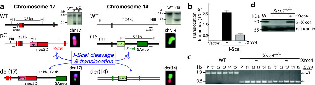
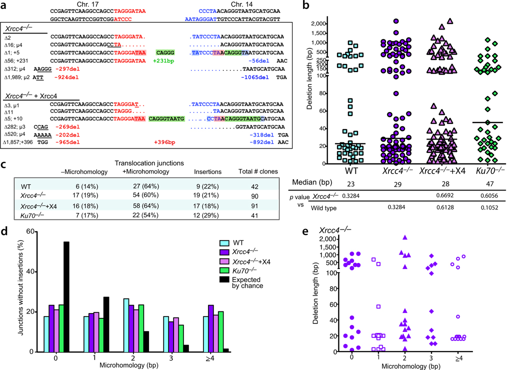
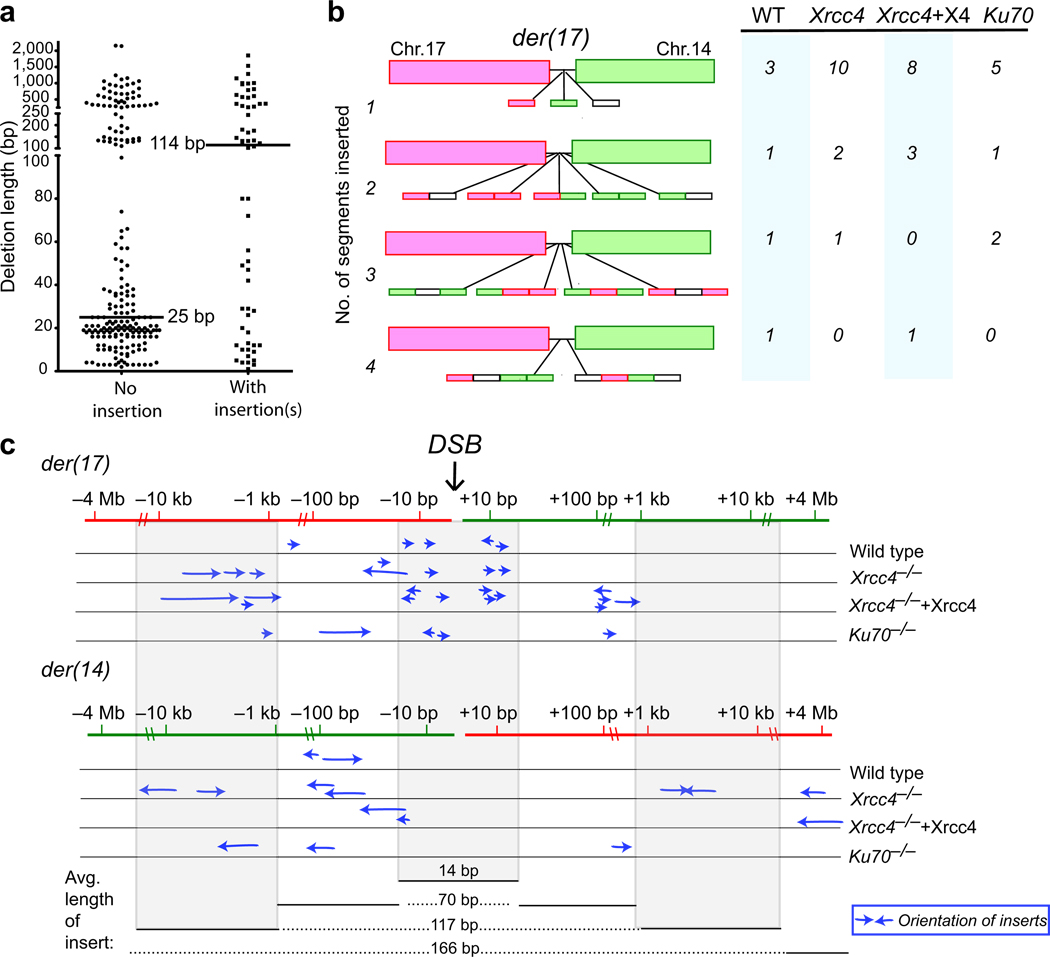
Chromosomal translocations in hematologic and mesenchymal tumors form overwhelmingly by nonhomologous end-joining (NHEJ). Canonical NHEJ, essential for the repair of radiation-induced and some programmed double-strand breaks (DSBs), requires the Xrcc4-ligase IV complex. For other DSBs, the requirement for Xrcc4-ligase IV is less stringent, suggesting the existence of alternative end-joining (alt-NHEJ) pathways. To understand the contributions of the canonical NHEJ and alt-NHEJ pathways, we examined translocation formation in cells deficient in Xrcc4-ligase IV. We found that Xrcc4-ligase IV is not required for but rather suppresses translocations. Translocation breakpoint junctions have similar characteristics in wild-type cells and cells deficient in Xrcc4-ligase IV, including an unchanged bias toward microhomology, unlike what is observed for intrachromosomal DSB repair. Complex insertions in some junctions show that joining can be iterative, encompassing successive processing steps before joining. Our results imply that alt-NHEJ is the primary mediator of translocation formation in mammalian cells.


Figures

Figure 1
Figure 1
Chromosomal translocations are suppressed by XRCC4/ligase IV. (a) Translocation reporter in Xrcc4−/− pCr15 cells. DSB formation on chromosomes 17 and 14 at the I-SceI sites, followed by interchromosomal NHEJ, results in a chromosomal translocation with a neo+ gene on der(17). FISH analysis indicates that parental pCr15 cells have normal chromosomes 17 (red) and 14 (green), whereas neo+ clones have derivative chromosomes. Vertical red bars are exons 1–5 from the targeted Pim1 locus on chr.17, and the vertical green bar is exon 20 from the Rb locus. Probes are located outside the targeting arms. HII, HincII; HIII, HindIII. (b) Translocation frequency is significantly increased in Xrcc4−/− cells, but is suppressed by Xrcc4 expression. (c) Confirmation of the genotype of the endogenous Xrcc4 alleles in wild-type (WT), Xrcc4−/− and Xrcc4-complemented cells. P, parental pCr15 cells of the indicated genotypes; t, neo+ translocation clones. (d) Western blot analysis demonstrating that transient expression of Xrcc4 restores wild-type Xrcc4 protein levels to Xrcc4−/− cells.
Figure 2
Figure 2
Translocation breakpoint junctions have similar characteristics in wild-type, Xrcc4−/− mutant and complemented cells, and Ku70−/− cells. (a) Representative der(17) translocation junction sequences obtained from Xrcc4−/− and Xrcc4-complemented cells. DNA ends generated by I-SceI on chrs.17 and 14 are indicated in red and blue, respectively. A summary of the various end modifications is presented to the left of each junction in bp: Δ, total deletion; µ, microhomology; +, insertion. Sequences are annotated as follows: del, deletion length from the DNA end; underline, microhomology; +, length of long insertion. The middle green sequences are short insertions from chr. 14; considering a template model for their insertion, the sequences in red shading (TAA) would be microhomology between the DNA ends that could anneal to act as a primer and the blue shading would represent microhomology for annealing after DNA synthesis between the 2 DNA ends (see text). (b) Deletion lengths for der(17) breakpoint junctions. Each value represents the combined deletion from both ends of an individual junction. The median deletion length for each genotype is indicated by a bar on the graph and the value is give below the graph. Deletion lengths do not differ significantly from each other (two-tailed Mann-Whitney test). +X4, transient complementation with Xrcc4. (c) Microhomology and insertion frequencies are similar for the four genotypes. (d) Distribution of microhomology lengths for der(17) breakpoint junctions. Only junctions with simple deletions (i.e., without an insertion) are included. (e) Lack of correlation between deletion length and microhomology use. Only junctions for Xrcc4−/− cells are plotted.
Figure 3
Figure 3
Insertions at breakpoint junctions display complex characteristics. (a) Distribution of deletion lengths for der(17) breakpoint junctions without and with insertions. Data are combined from all four genotypes; the deletion distributions for individual genotypes are shown in Supplementary Fig. 3d. (b) Der(17) insertions are comprised of one or more distinct segments of DNA. Most insertions at the der(17) breakpoint junctions are derived from one DNA segment, although others are comprised of up to 4 segments. Most inserted segments are derived from chr. 17 (red boxes) or chr. 14 (green boxes), although some of the inserted segments are derived from other sources or are too short to be mapped precisely (white boxes). Included in this analysis are all inserted sequences >4 bp. (c) Derivation of der(17) inserts. Each arrow represents the derivation of a segment inserted at a der(17) translocation breakpoint junction and the relative orientation of the insert relative to the centromere. Inserted segments are derived from sequences adjacent to a DSB or as far away as 4 Mb; those from nearby a DSB are generally short (≤20 bp), while more distantly-derived segments tend to be larger (up to 396 bp). Note that the inserted segments can be derived from sequences used to form either der(17) or der(14). The der(17) sequences from −1.5 kb to +1.2 kb comprise the neo intron (Fig. 1a).

Comment in

References

    1. van Gent DC, van der Burg M. Non-homologous end-joining, a sticky affair. Oncogene. 2007;26:7731–7740. - PubMed
    1. Jeggo PA. Identification of genes involved in repair of DNA double-strand breaks in mammalian cells. Radiat Res. 1998;150:S80–S91. - PubMed
    1. Lieber MR, Ma Y, Pannicke U, Schwarz K. Mechanism and regulation of human non-homologous DNA end-joining. Nat Rev Mol Cell Biol. 2003;4:712–720. - PubMed
    1. Chaudhuri J, et al. Evolution of the immunoglobulin heavy chain class switch recombination mechanism. Adv Immunol. 2007;94:157–214. - PubMed
    1. Greaves MF, Wiemels J. Origins of chromosome translocations in childhood leukaemia. Nat Rev Cancer. 2003;3:639–649. - PubMed

Publication types