. 2010 Apr;17(4):438-44.
doi: 10.1038/nsmb.1772. Epub 2010 Mar 7.

Structural basis for receptor recognition by New World hemorrhagic fever arenaviruses

Affiliations

Structural basis for receptor recognition by New World hemorrhagic fever arenaviruses

Jonathan Abraham et al. Nat Struct Mol Biol. 2010 Apr.

Abstract

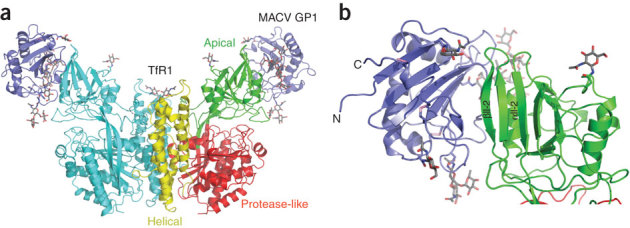
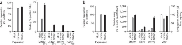
New World hemorrhagic fever arenaviruses are rodent-borne agents that cause severe human disease. The GP1 subunit of the surface glycoprotein mediates cell attachment through transferrin receptor 1 (TfR1). We report the structure of Machupo virus (MACV) GP1 bound with human TfR1. Atomic details of the GP1-TfR1 interface clarify the importance of TfR1 residues implicated in New World arenavirus host specificity. Analysis of sequence variation among New World arenavirus GP1s and their host-species receptors, in light of the molecular structure, indicates determinants of viral zoonotic transmission. Infectivities of pseudoviruses in cells expressing mutated TfR1 confirm that contacts at the tip of the TfR1 apical domain determine the capacity of human TfR1 to mediate infection by particular New World arenaviruses. We propose that New World arenaviruses that are pathogenic to humans fortuitously acquired affinity for human TfR1 during adaptation to TfR1 of their natural hosts.


Conflict of interest statement

The authors declare no competing financial interests.

Figures

Figure 1
Figure 1. Structure of the MACV GP1–human TfR1 complex.
(a) The butterfly-shaped TfR1 dimer is shown oriented with the cell surface at the bottom. One TfR1 protomer is colored according to the TfR1 domains: the apical domain is green, the protease-like domain is red and the helical domain is yellow. The other protomer is in cyan. MACV GP1, shown in purple, interacts with the TfR1 apical domain. The N-linked glycans are in gray. (b) Enlargement of the TfR1 apical domain with bound MACV GP1. Four N-linked glycans are on surfaces of the viral glycoprotein not involved in binding to TfR1. The N and C termini of MACV GP1 and the αII-2 helix and βII-2 strand of TfR1 are labeled.
Figure 2
Figure 2. MACV GP1.
(a) Domain organization of the MACV envelope glycoprotein (GP). GP is processed during its maturation into three noncovalently associated subunits: the stable signal peptide (SSP), GP1 and GP2. Sites of potential N-linked glycosylation are shown as tree diagrams. The triangle indicates the subtilisin/kexin isozyme 1 (SKI-1) cleavage site. TM, transmembrane segment. (b) Sequence alignment of MACV GP1 residues 80–250 with the GP1 proteins of NW clade B arenaviruses JUNV, TCRV, GTOV, AMAV and SABV. Secondary structure elements are shown for Residues are colored according to the ClustalX color scheme. GP1 residues that interact with hTfR1 are labeled with asterisks colored according to the interaction motifs shown in Figure 3; green for motif 1, magenta for motif 2, red for motif 3, yellow for motif 4 and blue for motif 5. (c) Ribbon diagram of The first N-acetylglucosamine is shown for glycosylation sites at Asn95, Asn137, Asn166 and Asn178. The four disulfide bonds that rigidify the GP1 core are in pink. The conserved Cys92-Cys237 and Cys135-Cys164 disulfide bridges are labeled with asterisks. The three loops that protrude from the GP1 core to contact TfR1 (L3, L7 and L10) are in yellow. Note that L10 is much shorter in the other viruses and will not contact TfR1.
Figure 3
Figure 3. Five interaction motifs in the MACV GP1-human TfR1 interface.
(a) Motif 1, centered on Tyr211 on the βII-2 strand in the receptor. (b) Motif 2, centered on TfR1 Asn348, which caps the C terminus of αII-2 helix. (c) Motif 3, centered on TfR1 Val210 in the βII-2 strand of the receptor, on the side opposite Tyr211. A segment unique to MACV GP1 is in red, with the Cys220-Cys229 disulfide bond in yellow. (d) Motif 4, at the lateral base of the TfR1 apical domain. An extensive network of polar interactions radiates from TfR1 Lys344. (e) Motif 5 at the lateral tip of the apical domain, centered on TfR1 Leu209. TfR1 residues Asp204 and Arg208, also shown, have been implicated previously in the host specificity of NW arenaviruses,. Asn95 is a conserved glycosylation site in New World arenavirus GP1s. The anchor residues for each motif are labeled with an asterisk. Hydrogen bonds are indicated as dashed lines.
Figure 4
Figure 4. Conservation of the GP1-TfR1 interface.
(a) Ribbon diagram of MACV GP1, as seen from the apical domain of TfR1 in the complex. GP1 residues that contact TfR1 are colored according to the interaction motifs shown in Figure 3, using the color scheme outlined in the caption to Figure 2b. (b) List of MACV GP1 residues that contact TfR1, colored according to the five interaction motifs. Residues found in analogous positions for the New World clade B arenaviruses JUNV, TCRV, GTOV, AMAV and SABV (based on the sequence alignment shown in Fig. 2b) are also shown. (c) Side-view ribbon diagram of the TfR1 apical domain. TfR1 residues that contact MACV GP1 are colored according to the five interaction motifs as in a. (d) List of hTfR1 residues that contact MACV GP1, colored according to the five interaction motifs. Residues found in analogous positions in C. callosus (MACV host), C. musculinus (JUNV host), A. jamaicensis (TCRV host), Z. brevicauda (GTOV host) and N. spinosus (AMAV host) TfR1 are listed based on sequence alignment (see Supplementary Fig. 3).
Figure 5
Figure 5. Importance of the tip of the apical domain in determining the role of human TfR1 as a receptor for pathogenic New World arenaviruses.
(a) CHO cells were transfected with vector alone (mock) or plasmids encoding hTfR1 (human), or hTfR1 with the Arg208Gly mutation (R208G); 48 h later, the cells were treated with an anti-Flag antibody to measure cell surface expression of receptors (left), or with immunoglobulin (Ig) fusion proteins containing the truncated GP1 subunits (GP1Δ) of MACV, JUNV or GTOV or the receptor binding domain of the severe acute respiratory syndrome coronavirus (SARS-CoV) spike protein (right). Association of these proteins with cells was measured by flow cytometry. (b) CHO cells were transfected with the indicated plasmids, and cell surface expression of the receptors (left) determined 48 h later with an anti-Flag antibody as in (a). Aliquots of these cells were infected in parallel with MACV, JUNV, GTOV or vesicular stomatitis virus (VSV) pseudoviruses expressing GFP. Infection levels were assessed 48 h later by measuring GFP expression by flow cytometry. Error bars indicate s.d.

Similar articles

Cited by

References

    1. Oldstone MB. Arenaviruses. I. The epidemiology molecular and cell biology of arenaviruses. Introduction. Curr. Top. Microbiol. Immunol. 2002;262:V–XII. - PubMed
    1. Bowen MD, Peters CJ, Nichol ST. The phylogeny of New World (Tacaribe complex) arenaviruses. Virology. 1996;219:285–290. doi: 10.1006/viro.1996.0248. - DOI - PubMed
    1. Clegg JC. Molecular phylogeny of the arenaviruses. Curr. Top. Microbiol. Immunol. 2002;262:1–24. - PubMed
    1. Emonet S, Lemasson JJ, Gonzalez JP, de Lamballerie X, Charrel RN. Phylogeny and evolution of old world arenaviruses. Virology. 2006;350:251–257. doi: 10.1016/j.virol.2006.01.026. - DOI - PubMed
    1. Charrel RN, de Lamballerie X. Arenaviruses other than Lassa virus. Antiviral Res. 2003;57:89–100. doi: 10.1016/S0166-3542(02)00202-4. - DOI - PubMed

Publication types

MeSH terms

Associated data

LinkOut - more resources