. 2010 May 27;29(21):3146-52.
doi: 10.1038/onc.2010.52. Epub 2010 Mar 8.

Activation of EGFR by proteasome inhibition requires HB-EGF in pancreatic cancer cells

Affiliations

Activation of EGFR by proteasome inhibition requires HB-EGF in pancreatic cancer cells

C M Sloss et al. Oncogene. .

Abstract

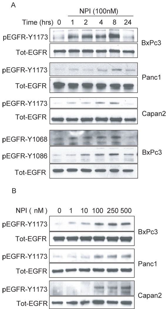
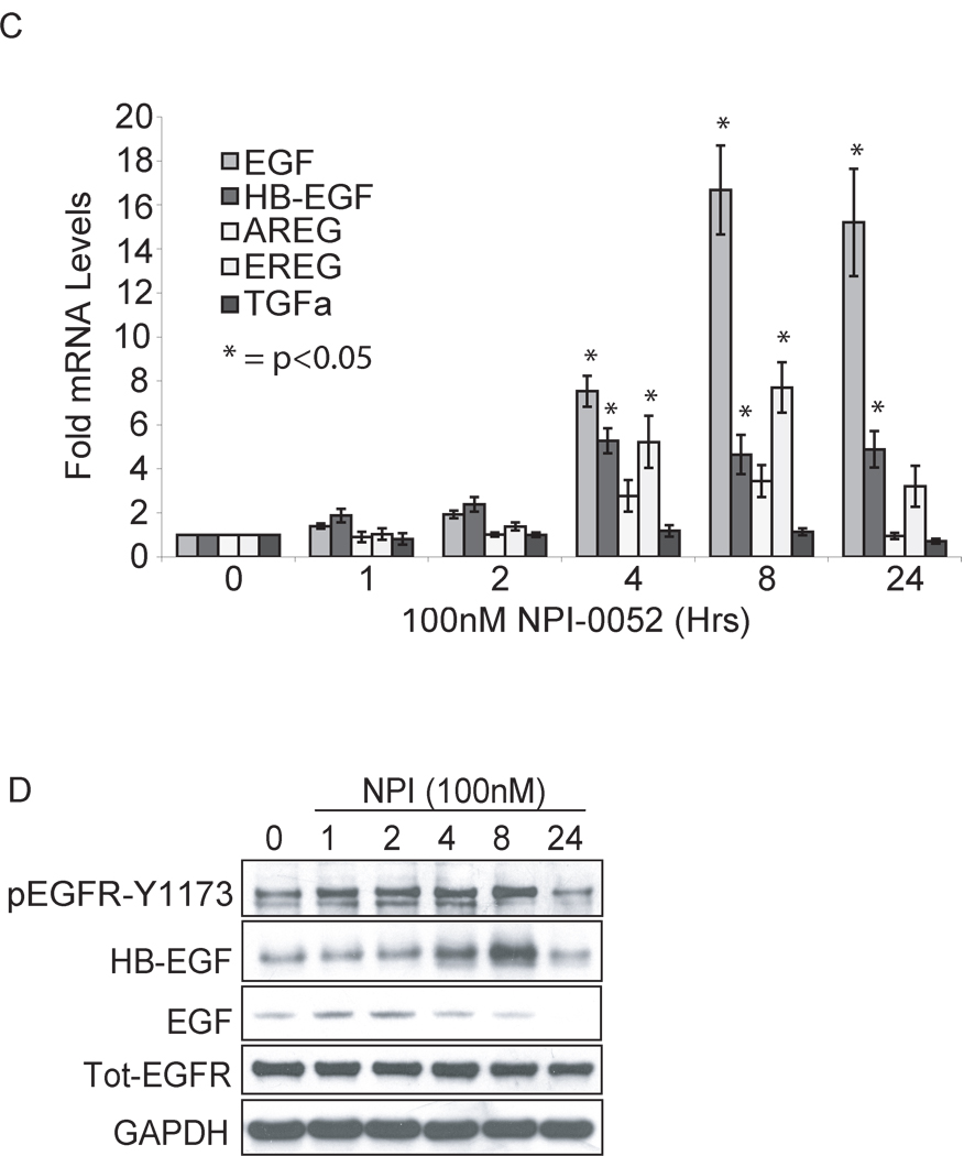
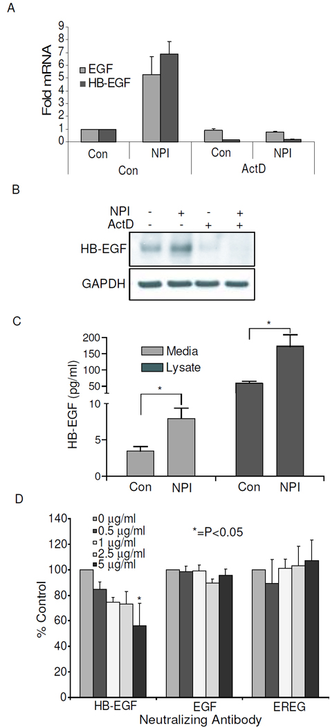
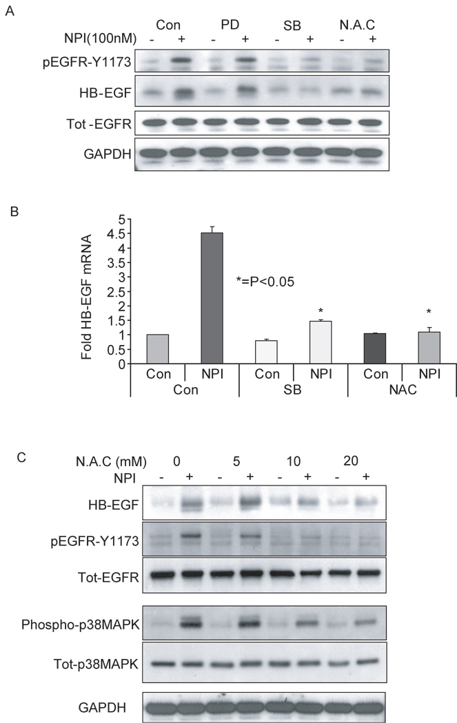
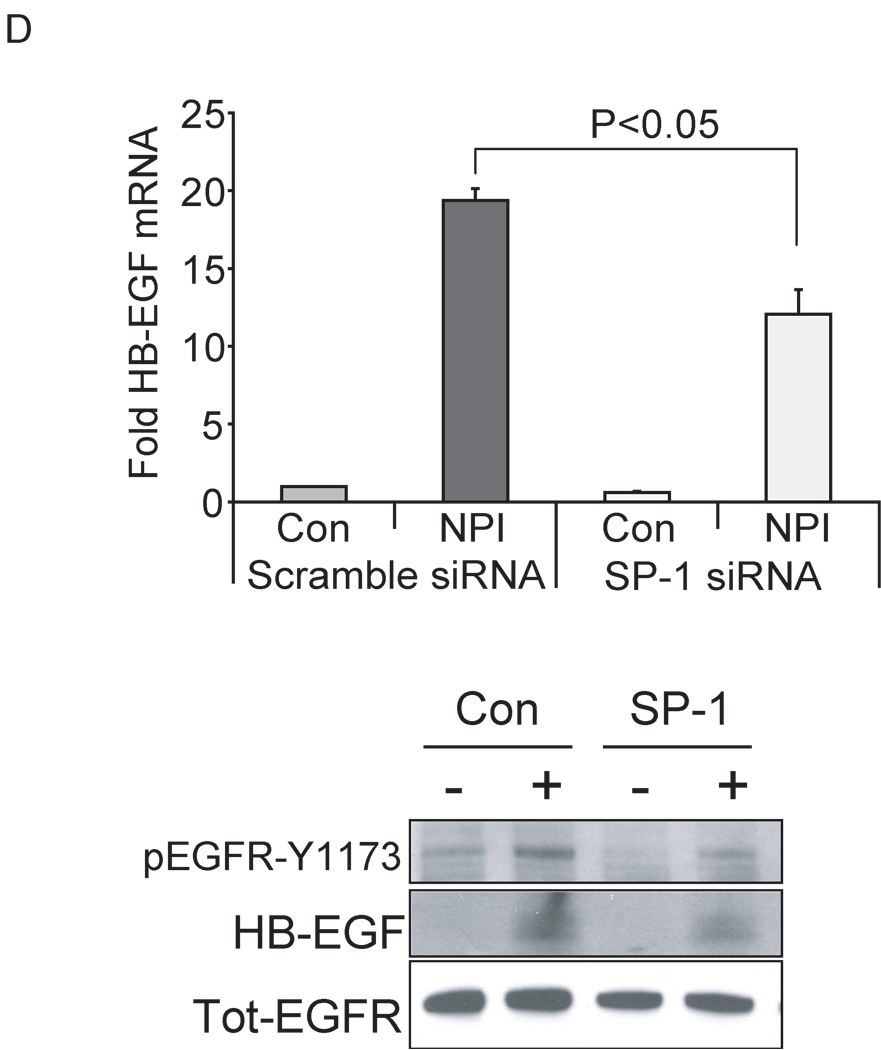
Resistance to drug treatments underlies the high lethality of pancreatic ductal adenocarcinoma. Along with others, we have recently identified that proteasome inhibition is a promising therapeutic option in this highly refractory disease. The pleiotropic effects of proteasome inhibition include the activation of apoptotic signaling pathways and also antiapoptotic signaling pathways such as EGFR, AKT and the MAP kinases that reduce the apoptotic potential of this class of drug. In this study, we sought to determine the mechanism behind the activation of EGFR in response to proteasome inhibition in pancreatic cancer cells. We found that the second-generation proteasome inhibitor NPI-0052 induced the mRNA transcription of several EGFR family ligands (EGF, HB-EGF and epiregulin), however only increases in HB-EGF were detected at the protein level. Using both pharmacological inhibitors and lentiviral-mediated shRNA knockdown of EGFR ligand expression, we discovered that ligand cleavage by MMP/ADAMs and HB-EGF expression is required for activation of EGFR in response to proteasome inhibition. Furthermore, we discover that induction of HB-EGF is dependent on reactive oxygen species and p38-MAPK signaling but not ERK and that the transcription factor SP-1 is involved in NPI-0052-induced HB-EGF transcription. Together, these results indicate that stress signaling leading to induction of HB-EGF expression and increases in MMP/ADAM-dependent HB-EGF cleavage are responsible for proteasome inhibitor-induced activation of EGFR in pancreatic cancer cells.


Figures

Figure 1
Figure 1
NPI-0052 induces EGFR activation in a time and dose dependant manner and induces HER family ligand expression. BxPC3, Panc1, Capan2 were obtained from the ATCC (Rockville, MD). Capan2 and Panc1 cells were grown in DMEM; BxPC3 in RPMI. Media were supplemented with 10% fetal bovine serum and 100µg/ml penicillin & streptomycin. (A) BxPc3, Panc1 and Capan2 cells were treated with 100nM NPI-0052 (Nereus Pharmaceticals, CA) for the indicated times or (B) with the indicated concentrations of NPI-0052 for 4 hours. Whole cell extracts were prepared in RIPA buffer (50mM Tris-HCl pH 7.4, 150mM NaCl, 1mM EDTA, 1% Triton x-100, 1% Sodium Deoxycholate, 0.1% SDS, 1µg/ml Aprotinin, 1µg/ml Leupeptin, 1µg/ml Pepstatin, 1mM Na3VO4, 50mM b-Glycerophosphate, 1mM PMSF) and immunoblotted for the indicated pTyr-residues on EGFR or the total form of EGFR. (C) BxPc3 cells were treated with 100nM NPI-0052 for the indicated times. Total RNA was isolated using the RNeasy mini kit (Qiagen, Valencia, CA) and 250ng of RNA was used to measure the relative expression of EGFR ligands by RT-qPCR analysis using the indicated hydrolysis probes purchased from Applied Biosystem (Foster City, CA) in 50µl reactions performed using one-step-qRT-PCR (Invitrogen, Carlsbad, CA) on an ABI7300 system. GAPDH was used as control. Error bars represent standard error of the mean (S.E.M) of at least 3 separate experiments, *=P<0.05 by one-way-ANOVA with Dunnet’s post test relative to untreated cells. (D) BxPc3 cells were treated with 100nM NPI-0052 for the indicated times and expression of the indicated proteins measured by immunoblotting.
Figure 1
Figure 1
NPI-0052 induces EGFR activation in a time and dose dependant manner and induces HER family ligand expression. BxPC3, Panc1, Capan2 were obtained from the ATCC (Rockville, MD). Capan2 and Panc1 cells were grown in DMEM; BxPC3 in RPMI. Media were supplemented with 10% fetal bovine serum and 100µg/ml penicillin & streptomycin. (A) BxPc3, Panc1 and Capan2 cells were treated with 100nM NPI-0052 (Nereus Pharmaceticals, CA) for the indicated times or (B) with the indicated concentrations of NPI-0052 for 4 hours. Whole cell extracts were prepared in RIPA buffer (50mM Tris-HCl pH 7.4, 150mM NaCl, 1mM EDTA, 1% Triton x-100, 1% Sodium Deoxycholate, 0.1% SDS, 1µg/ml Aprotinin, 1µg/ml Leupeptin, 1µg/ml Pepstatin, 1mM Na3VO4, 50mM b-Glycerophosphate, 1mM PMSF) and immunoblotted for the indicated pTyr-residues on EGFR or the total form of EGFR. (C) BxPc3 cells were treated with 100nM NPI-0052 for the indicated times. Total RNA was isolated using the RNeasy mini kit (Qiagen, Valencia, CA) and 250ng of RNA was used to measure the relative expression of EGFR ligands by RT-qPCR analysis using the indicated hydrolysis probes purchased from Applied Biosystem (Foster City, CA) in 50µl reactions performed using one-step-qRT-PCR (Invitrogen, Carlsbad, CA) on an ABI7300 system. GAPDH was used as control. Error bars represent standard error of the mean (S.E.M) of at least 3 separate experiments, *=P<0.05 by one-way-ANOVA with Dunnet’s post test relative to untreated cells. (D) BxPc3 cells were treated with 100nM NPI-0052 for the indicated times and expression of the indicated proteins measured by immunoblotting.
Figure 2
Figure 2
Ligand expression is actinomycin-D sensitive. BxPc3 cells were treated with vehicle or 100nM NPI-0052 in the presence or absence of 5µg/ml ActinomycinD (Sigma, St. Louis, MO) for 4 hours. The relative expressions of HB-EGF and EGF were measured by (A) RT-qPCR or (B) immunoblotting. NPI-0052 treatment induces HB-EGF shedding. (C) BxPc3 cells were treated for 8 hours with vehicle or 100nM NPI-0052, growth media and whole cell extracts were analyzed for levels of HB-EGF expression by ELISA using antibodies purchased from R&D systems (Minneapolis, MN). Samples were pre-cleared by centrifugation at 5000xg for 5mins at 4°C and 100µl/well used for analysis as described in the manufacturers protocol and as described previously (Wang et al., 2007a). A standard curve was generated using rh-HB-EGF (R&D) and used to quantify unknowns. Error bars represent S.E.M of at least 3 separate experiments. * indicates P<0.05 by students t-test. (D) BxPc3 cells were treated for 8 hours with vehicle or 100nM NPI-0052 in the presence of the indicated amounts of neutralizing antibodies (R&D systems). Lysates were analysed for activation of EGFR by immunoblot. Histogram represents mean quantification by scanning densitometry of at least 3 separate immunoblot bands (representative blots are shown in supplementary figure 2A). Error bars represent S.E.M. *=P<0.05 by one-way-ANOVA with Dunnet’s post test relative to NPI treated cells with 0µg/ml of neutralizing antibody.
Figure 3
Figure 3
Activation of EGFR is MMP/ADAM and HB-EGF dependant. BxPc3 cells were treated for 8 hours with vehicle (−) or 100nM NPI-0052 (+) with either (A) the indicated concentrations of the MMP/ADAM inhibitor GM6001 (Sigma) or (B) the indicated concentrations of the HB-EGF inhibitor crm-197 (EMD biosciences, Madison, WI) and expression of the indicated proteins measured by immunoblotting. Knockdown of HB-EGF but not EGF or epiregulin prevents EGFR activation. Control Plko1 vector, EGF (NM_001963.2-2240s1c1); HB-EGF (NM_001945.1-827s1c1) or EREG (NM_001432.1-341s1c1) (Sigma) were used to produce lentiviral particles in 293T cells as described in the manufacturers protocol. After lentiviral transduction, stable BxPC3 clones were generated by passaging cells in 5µg/ml Puromycin (Fisher, Waltham, MA). Control cells (Bx-Plko1) or cells lacking HB-EGF (Bx-shHB), EGF (Bx-shEGF) or epiregulin (Bx-shEREG) were treated with vehicle or 100nM NPI-0052 for 8 hours and (C) the relative expression of ligands were measured using RT-qPCR (error bars represent S.E.M, P values represent analysis by student’s t-test), or (D) cell lysates were immunoblotted for the indicated proteins.
Figure 4
Figure 4
HB-EGF induction and EGFR activation is dependent on ROS and p38-MAPK but not ERK. (A) BxPc3 cells were treated for 8hrs with vehicle (−) or 100nM NPI-0052 (+) with inhibitors of the ERK pathway (10µM PD98059); the p38 MAPK pathway (2.5µM SB203580) or ROS (10mM NAC). Cell lysates were analyzed for the indicated proteins by immunoblot or (B) for HB-EGF mRNA levels by RT-qPCR. Error bars represent S.E.M of at least 2 experiments. *=P<0.05 by students t-test vs NPI treated control. (C) BxPc3 cells were treated for 8hrs with vehicle (−) or 100nM NPI-0052 (+) with the indicated concentrations of NAC. Cell lysates were analyzed for the indicated proteins by immunoblot. (D) Panc1 cells in 6 well plates were transfected with 50nM of either Scrambled or anti-SP-1 stealth-siRNA (Invitrogen), using 5µl of RNAiMax (Invitrogen), in 500µl of Optimem (Invitrogen), added to a final volume of 3ml of antibiotic free media per well as described in the manufacturers protocol. After 48 hours cell lysates were analyzed for relative HB-EGF mRNA levels by RT-qPCR or by immunoblot for the indicated proteins. Error bars represent S.E.M of at least 3 experiments. P<0.05 by students t-test.
Figure 4
Figure 4
HB-EGF induction and EGFR activation is dependent on ROS and p38-MAPK but not ERK. (A) BxPc3 cells were treated for 8hrs with vehicle (−) or 100nM NPI-0052 (+) with inhibitors of the ERK pathway (10µM PD98059); the p38 MAPK pathway (2.5µM SB203580) or ROS (10mM NAC). Cell lysates were analyzed for the indicated proteins by immunoblot or (B) for HB-EGF mRNA levels by RT-qPCR. Error bars represent S.E.M of at least 2 experiments. *=P<0.05 by students t-test vs NPI treated control. (C) BxPc3 cells were treated for 8hrs with vehicle (−) or 100nM NPI-0052 (+) with the indicated concentrations of NAC. Cell lysates were analyzed for the indicated proteins by immunoblot. (D) Panc1 cells in 6 well plates were transfected with 50nM of either Scrambled or anti-SP-1 stealth-siRNA (Invitrogen), using 5µl of RNAiMax (Invitrogen), in 500µl of Optimem (Invitrogen), added to a final volume of 3ml of antibiotic free media per well as described in the manufacturers protocol. After 48 hours cell lysates were analyzed for relative HB-EGF mRNA levels by RT-qPCR or by immunoblot for the indicated proteins. Error bars represent S.E.M of at least 3 experiments. P<0.05 by students t-test.

Similar articles

Cited by

References

    1. Blaine SA, Ray KC, Branch KM, Robinson PS, Whitehead RH, Means AL. The epidermal growth factor receptor regulates pancreatic fibrosis. Am J Physiol Gastrointest Liver Physiol. 2009 - PMC - PubMed
    1. Buck E, Eyzaguirre A, Haley JD, Gibson NW, Cagnoni P, Iwata KK. Inactivation of Akt by the epidermal growth factor receptor inhibitor erlotinib is mediated by HER-3 in pancreatic and colorectal tumor cell lines and contributes to erlotinib sensitivity. Mol Cancer Ther. 2006;5:2051–2059. - PubMed
    1. Codony-Servat J, Tapia MA, Bosch M, Oliva C, Domingo-Domenech J, Mellado B, et al. Differential cellular and molecular effects of bortezomib, a proteasome inhibitor, in human breast cancer cells. Mol Cancer Ther. 2006;5:665–675. - PubMed
    1. D'Addario M, Arora PD, McCulloch CA. Role of p38 in stress activation of Sp1. Gene. 2006;379:51–61. - PubMed
    1. Edwards JP, Zhang X, Mosser DM. The expression of heparin-binding epidermal growth factor-like growth factor by regulatory macrophages. J Immunol. 2009;182:1929–1939. - PMC - PubMed

Publication types

MeSH terms

Substances