. 2010 Feb 23;8(2):e1000315.
doi: 10.1371/journal.pbio.1000315.

Voltage-dependent gating in a "voltage sensor-less" ion channel

Affiliations

Voltage-dependent gating in a "voltage sensor-less" ion channel

Harley T Kurata et al. PLoS Biol. .

Abstract

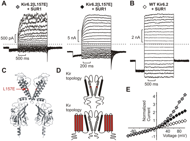
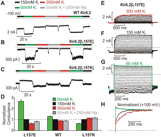
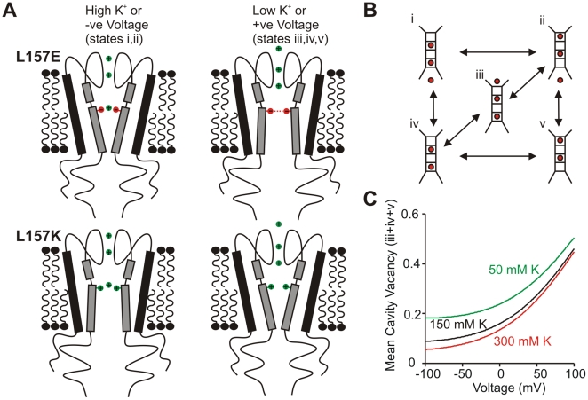
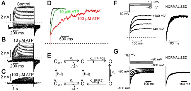
The voltage sensitivity of voltage-gated cation channels is primarily attributed to conformational changes of a four transmembrane segment voltage-sensing domain, conserved across many levels of biological complexity. We have identified a remarkable point mutation that confers significant voltage dependence to Kir6.2, a ligand-gated channel that lacks any canonical voltage-sensing domain. Similar to voltage-dependent Kv channels, the Kir6.2[L157E] mutant exhibits time-dependent activation upon membrane depolarization, resulting in an outwardly rectifying current-voltage relationship. This voltage dependence is convergent with the intrinsic ligand-dependent gating mechanisms of Kir6.2, since increasing the membrane PIP2 content saturates Po and eliminates voltage dependence, whereas voltage activation is more dramatic when channel Po is reduced by application of ATP or poly-lysine. These experiments thus demonstrate an inherent voltage dependence of gating in a "ligand-gated" K+ channel, and thereby provide a new view of voltage-dependent gating mechanisms in ion channels. Most interestingly, the voltage- and ligand-dependent gating of Kir6.2[L157E] is highly sensitive to intracellular [K+], indicating an interaction between ion permeation and gating. While these two key features of channel function are classically dealt with separately, the results provide a framework for understanding their interaction, which is likely to be a general, if latent, feature of the superfamily of cation channels.


Conflict of interest statement

The authors have declared that no competing interests exist.

Figures

Figure 1
Figure 1. Voltage-dependent activation of Kir6.2[L157E] channels.
(A,B) Representative inside-out patch clamp recordings from (A) two different Kir6.2[L157E] membrane patches and (B) a WT Kir6.2 membrane patch (both co-expressed with SUR1). Patches were pulsed to voltages between −100 and +100 mV, with a holding potential of −50 mV. (C) Molecular model of Kir6.2, with residue 157 highlighted in red. (D) Transmembrane topologies of Kir and Kv channel families, with elements underlying voltage-dependent gating colored red in each case. (E) Current-voltage relationships illustrating outward rectification of Kir6.2[L157E] channels. Symbols correspond to the recordings depicted in panels in A–B.
Figure 2
Figure 2. Voltage-dependent gating of Kir6.2[L157E] channels interacts with PIP2-regulated open probability.
(A–D) Representative current traces from a Kir6.2[L157E] membrane patch, (A) immediately after excision, (B) in 1 mM ATP, (C) after exposure to 5 µg/mL PIP2, (D) after brief exposure to the PIP2 antagonist poly-lysine. (E) Currents from A, C, and D are normalized to peak to illustrate the effects of basal open probability (determined by PIP2) on activation kinetics and on the activating fraction of peak current. (F,G) Compiled data from 4 Kir6.2[L157E] membrane patches, illustrating the relationship between ATP sensitivity (an index of open state stability), and (F) activation kinetics or (G) activating fraction. At higher open state stability, a smaller fraction of the peak current exhibits time-dependent activation, and the kinetics of activation are markedly faster. In (F,G), data are presented from four patches, with each symbol type reflecting a different membrane patch. Dashed lines are linear regression fits to each individual patch, while the solid line is a fit to the entire data set.
Figure 3
Figure 3. Kinetic model describing voltage-dependent activation of Kir6.2[L157E] over a range of voltage and basal open probability.
(A) Current records collected after saturating open probability with PIP2 (i), followed by progressive reduction of open state stability with brief applications of poly-lysine (ii–v). In the right-hand panel, steady-state currents were normalized to fully activated currents (record i) to illustrate the extent of activation at each voltage. (B) Kinetic model depicting two tiers of gating—a low Po tier (lower) and a high Po tier (upper). In the high Po tier, the KCO equilibrium constant is 7-fold larger. Equilibria between the high and low Po tiers are governed by the Kv constant, and g is a factor included to preserve reversibility (g = KCO*/KCO).
Figure 4
Figure 4. Depolarization increases open probability of Kir6.2[L157E] channels.
(A) Current records from membrane patches containing few channels (likely three per patch) for Kir6.2[L157E] or WT Kir6.2 recorded in symmetrical 150 mM K+ conditions. (B) Single-channel currents between −100 and +100 mV in WT Kir6.2 and Kir6.2[L157E] channels. The L157E mutation has no significant effect on single channel conductance. (C) Open probability of Kir6.2[L157E] (top) or WT Kir6.2 (bottom) channels measured from membrane patches containing 1−5 channels, between −100 and +100 mV (n = 3 for WT and 4 for L157E).
Figure 5
Figure 5. Position 157 affects the internal K+ sensitivity of Kir6.2.
(A–C) Continuous current records at −100 mV depicting responses to altered internal ionic conditions in inside-out membrane patches expressing (A) WT Kir6.2, (B) Kir6.2[L157E], or (C) Kir6.2[L157K]. The L157E mutation exaggerates the response observed in WT Kir6.2, while the L157K mutation reverses the WT response to intracellular K+. (D) Using voltage-step protocols, the chord conductance between −100 and −80 mV was calculated in all Kint conditions and normalized to the conductance in 150 mM K+ in each patch (n = 29 for WT Kir6.2, 19 for Kir6.2[L157E], and 20 for Kir6.2[L157K]). (E–G) Current records from a Kir6.2[L157E] inside-out patch, at voltages from −100 to +100 mV. (H) Currents elicited by a step to +100 mV, normalized to peak current, in the ionic conditions depicted in panels E–G.
Figure 6
Figure 6. Hypothetical mechanism of convergent regulation by voltage and internal ions.
(A) Cation occupancy in the cavity ion binding site will mitigate repulsion between glutamates substituted at position 157, favoring the closed state relative to conditions in which the cavity site is unoccupied. Introduction of a positive charge at position 157 would exhibit an opposite response to cavity site occupancy. (B) Permeation model, with boxes representing selectivity filter binding sites, flanked by an external binding site (top) and the cavity site (bottom). (C) Simulation of voltage and internal K+-dependent changes in mean occupancy of the cavity ion binding site (sum of probability of occupancy in states i+ii), using parameters generated to describe permeation through KcsA channels .
Figure 7
Figure 7. Predictions of gating coupled to changes in permeant ion occupancy.
(A–C) In the presence of internal ATP, which acts by prolonging interburst intervals, the kinetics of channel activation are markedly slowed. (D) Normalized current traces recorded at +100 mV, in control and various internal ATP concentrations. (E) Extension of the scheme in Figure 3B, including ATP binding to closed states. In this scheme, ATP stabilizes the channel closed state, thereby prolonging the interburst intervals. (F,G) Kinetics of activation (F) and deactivation (G) and normalized in right-hand panels to illustrate very weak voltage dependence of kinetics.
Figure 8
Figure 8. Ionic strength affects channel interactions with PIP2.
WT Kir6.2 channels were fully run-down in high Mg2+ and subjected to increasing concentrations of di-C8 PIP2 in either (A) 50 mM or (B) 300 mM internal K+. PIP2 results in more significant current recovery in low ionic strength conditions. Similar observations were made in six membrane patches.

Similar articles

Cited by

References

    1. Long S. B, Campbell E. B, MacKinnon R. Voltage sensor of Kv1.2: structural basis of electromechanical coupling. Science. 2005;309:903–908. - PubMed
    1. Ahern C. A, Horn R. Focused electric field across the voltage sensor of potassium channels. Neuron. 2005;48:25–29. - PubMed
    1. Bezanilla F. How membrane proteins sense voltage. Nat Rev Mol Cell Biol. 2008;9:323–332. - PubMed
    1. Grabe M, Lai H. C, Jain M, Jan Y. N, Jan L. Y. Structure prediction for the down state of a potassium channel voltage sensor. Nature. 2007;445:550–553. - PubMed
    1. Tombola F, Pathak M. M, Gorostiza P, Isacoff E. Y. The twisted ion-permeation pathway of a resting voltage-sensing domain. Nature. 2007;445:546–549. - PubMed

Publication types

MeSH terms