

Action Spectroscopy on Dense Samples of Photosynthetic Reaction Centers of Rhodobacter sphaeroides WT Based on Nanosecond Laser-Flash C Photo-CIDNP MAS NMR
- PMID: 20208980
- PMCID: PMC2826643
- DOI: 10.1007/s00723-009-0103-6
Action Spectroscopy on Dense Samples of Photosynthetic Reaction Centers of Rhodobacter sphaeroides WT Based on Nanosecond Laser-Flash C Photo-CIDNP MAS NMR
Abstract
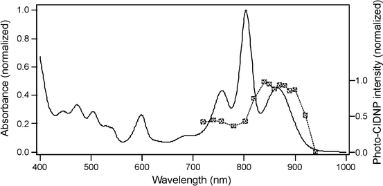
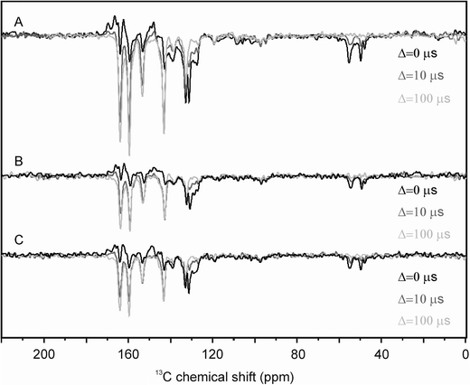
Photochemically induced dynamic nuclear polarization magic-angle spinning nuclear magnetic resonance (photo-CIDNP MAS NMR) allows for the investigation of the electronic structure of the photochemical machinery of photosynthetic reaction centers (RCs) at atomic resolution. For such experiments, either continuous radiation from white xenon lamps or green laser pulses are applied to optically dense samples. In order to explore their optical properties, optically thick samples of isolated and quinone-removed RCs of the purple bacteria of Rhodobacter sphaeroides wild type are studied by nanosecond laser-flash (13)C photo-CIDNP MAS NMR using excitation wavelengths between 720 and 940 nm. Action spectra of both the transient nuclear polarization as well as the nuclear hyperpolarization, remaining in the electronic ground state at the end of the photocycle, are obtained. It is shown that the signal intensity is limited by the amount of accessible RCs and that the different mechanisms of the photo-CIDNP production rely on the same photophysical origin, which is the photocycle induced by one single photon.
Figures


Similar articles
-
15N photo-CIDNP MAS NMR analysis of a bacterial photosynthetic reaction center of Rhodobacter sphaeroides wildtype.J Chem Phys. 2019 Nov 21;151(19):195101. doi: 10.1063/1.5128783. J Chem Phys. 2019. PMID: 31757137
-
Assignment of NMR resonances of protons covalently bound to photochemically active cofactors in photosynthetic reaction centers by 13C-1H photo-CIDNP MAS-J-HMQC experiment.J Magn Reson. 2019 Jan;298:64-76. doi: 10.1016/j.jmr.2018.11.013. Epub 2018 Nov 29. J Magn Reson. 2019. PMID: 30529893
-
Spectral editing through laser-flash excitation in two-dimensional photo-CIDNP MAS NMR experiments.J Magn Reson. 2014 Sep;246:9-17. doi: 10.1016/j.jmr.2014.06.007. Epub 2014 Jun 27. J Magn Reson. 2014. PMID: 25063951
-
Analysis of electron donors in photosystems in oxygenic photosynthesis by photo-CIDNP MAS NMR.J Photochem Photobiol B. 2015 Nov;152(Pt B):261-71. doi: 10.1016/j.jphotobiol.2015.08.001. Epub 2015 Aug 7. J Photochem Photobiol B. 2015. PMID: 26282679 Review.
-
Photo-CIDNP in Solid State.Appl Magn Reson. 2022;53(3-5):521-537. doi: 10.1007/s00723-021-01322-5. Epub 2021 Apr 6. Appl Magn Reson. 2022. PMID: 33840910 Free PMC article. Review.
Cited by
-
The electronic structure of the primary electron donor of reaction centers of purple bacteria at atomic resolution as observed by photo-CIDNP 13C NMR.Proc Natl Acad Sci U S A. 2009 Dec 29;106(52):22281-6. doi: 10.1073/pnas.0908608106. Epub 2009 Dec 11. Proc Natl Acad Sci U S A. 2009. PMID: 20018724 Free PMC article.
-
Tailored flavoproteins acting as light-driven spin machines pump nuclear hyperpolarization.Sci Rep. 2020 Oct 29;10(1):18658. doi: 10.1038/s41598-020-75627-z. Sci Rep. 2020. PMID: 33122681 Free PMC article.
-
N Photo-CIDNP MAS NMR To Reveal Functional Heterogeneity in Electron Donor of Different Plant Organisms.Appl Magn Reson. 2012 Feb;42(1):57-67. doi: 10.1007/s00723-011-0283-8. Epub 2011 Nov 15. Appl Magn Reson. 2012. PMID: 22303078 Free PMC article.
-
Nuclear spin-hyperpolarization generated in a flavoprotein under illumination: experimental field-dependence and theoretical level crossing analysis.Sci Rep. 2019 Dec 5;9(1):18436. doi: 10.1038/s41598-019-54671-4. Sci Rep. 2019. PMID: 31804538 Free PMC article.
-
Electronic Structures of Radical-Pair-Forming Cofactors in a Heliobacterial Reaction Center.Molecules. 2024 Feb 27;29(5):1021. doi: 10.3390/molecules29051021. Molecules. 2024. PMID: 38474533 Free PMC article.
References
-
- F. Haxo, Comparative Biochemistry of Photoreactive Systems (Academic Press, New York, 1960), pp. 339–376
-
- E.I. Rabinowitch, Govindjee, Photosynthesis (Wiley, New York, 1969), pp. 141–151
-
- K.M. Hartmann, in Biophysik, vol. 2, eds. by W. Hoppe, W. Lohmann, H. Markl, H. Ziegler (Springer-Verlag, Berlin, 1982), pp. 122–152
-
- L.O. Björn (ed.), Photobiology (Springer, New York, 2008)
-
- Vanginkel G, Kleinenhammans JW. Photochem. Photobiol. 1980;31:385–395. doi: 10.1111/j.1751-1097.1980.tb02557.x. - DOI
LinkOut - more resources
Full Text Sources