. 2010 Mar 1;5(3):e9470.
doi: 10.1371/journal.pone.0009470.

Molecular optical imaging with radioactive probes

Affiliations

Molecular optical imaging with radioactive probes

Hongguang Liu et al. PLoS One. .

Abstract

Background: Optical imaging (OI) techniques such as bioluminescence and fluorescence imaging have been widely used to track diseases in a non-invasive manner within living subjects. These techniques generally require bioluminescent and fluorescent probes. Here we demonstrate the feasibility of using radioactive probes for in vivo molecular OI.

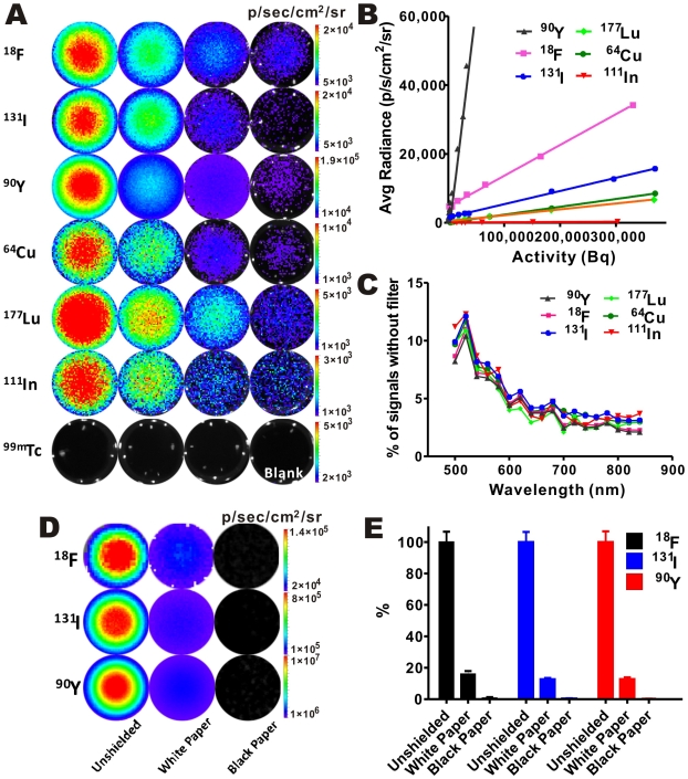
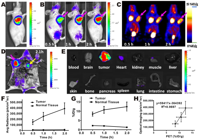
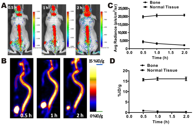
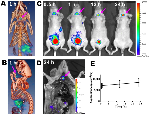
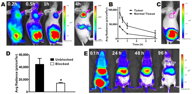
Methodology/principal findings: By taking the advantages of low energy window of light (1.2-3.1 eV, 400-1000 nm) resulting from radiation, radionuclides that emit charged particles such as beta(+) and beta(-) can be successfully imaged with an OI instrument. In vivo optical images can be obtained for several radioactive probes including 2-deoxy-2-[(18)F]fluoro-D-glucose ([(18)F]FDG), Na(18)F, Na(131)I, (90)YCl(3) and a (90)Y labeled peptide that specifically target tumors.

Conclusions/significance: These studies demonstrate generalizability of radioactive OI technique. It provides a new molecular imaging strategy and will likely have significant impact on both small animal and clinical imaging.


Conflict of interest statement

Competing Interests: The authors have declared that no competing interests exist.

Figures

Figure 1
Figure 1. Optical signals are detectable by OI instruments and have a continuous spectrum.
(A) Most nuclides, except 99mTc, provide OI signals with sensitivity within as low as 0.004–0.370 MBq (0.1–10 µCi) range. (18F: 5, 2, 1, 0.1 µCi; 131I: 10, 5, 1, 0.1 µCi; 90Y: 5, 1, 0.2, 0.01 µCi; 64Cu: 10, 5, 1, 0.1 µCi; 177Lu: 10, 5, 2, 0.5 µCi; 111In: 10, 5, 1, 0.5 µCi; 99mTc: 20, 10, 5 µCi ). (B) Detection sensitivity of different radionuclides. Radionuclides with a higher K value have stronger signal intensity. (C) Similar spectra were ovserved from different radionuclides. Results are presented as the ratio of photons detected using a narrow band emission filter (20nm bandwidth) versus total photons detected without a filter. (D, E) Shielding tests (D) and quantification analysis of imaging signals (E).
Figure 2
Figure 2. Phantom imaging studies with radioactive OI and PET.
(A–C) Radioactive OI of 18F, 131I and 90Y phantoms.
Figure 3
Figure 3. In vivo radioactive OI of [18F]FDG in comparison with microPET.
(A) Bioluminescence image of a nude mouse bearing C6-FLuc tumor. (B,C) Radioactive OI and microPET imaging of a nude mouse bearing C6-FLuc tumor injected via tail vein with [18F]FDG at 0.5, 1, 2 h p.i. (D,E) Radioactive OI of the mouse after opening the thorax (D) and exposure the organs (E) at 2.1 h p.i. (F–H) Quantitative analysis of radioactive OI (F) and microPET (G) results and their correlation (H).
Figure 4
Figure 4. Dual modality Na18F imaging.
Radioactive OI (A) and microPET imaging (B) of Na18F at 0.5, 1, 2 h after i.v. injection. Quantitative analysis of radioactive OI (C) and microPET (D) results.
Figure 5
Figure 5. In vivo radioactive OI of Na131I compared to SPECT/CT imaging.
Coronal (A) and sagittal (B) images of SPECT/CT imaging at 1 h after injection of Na131I probe. (C) Radioactive OI of a normal mouse at 0.5, 1, 12, 24 h after injection of Na131I via tail vein. (D) Radioactive OI of a normal mouse after opening the thorax 24 h post injection of Na131I. (E) Quantitative analysis (n = 3) of thyroid uptake of Na131I from radioactive OI results.
Figure 6
Figure 6. In vivo radioactive OI of 90Y-RGD-BBN and 90YCl3.
Radioactive OI (A) and quantitative analysis (n = 3) (B) of 90Y-RGD-BBN in mice bearing PC3 tumor. (C,D) Receptor blocking studies of 90Y-RGD-BBN probe (at 1 h post-injection) using radioactive OI (C) and their quantification analysis (n = 3) (D). (E) Radioactive OI of 90YCl3 at various time points p.i.

Similar articles

Cited by

References

    1. Massoud TF, Gambhir SS. Molecular imaging in living subjects: seeing fundamental biological processes in a new light. Genes Dev. 2003;17:545–580. - PubMed
    1. Gambhir SS. Molecular imaging of cancer with positron emission tomography. Nat Rev Cancer. 2002;2:683–693. - PubMed
    1. Weissleder R. Molecular imaging in cancer. Science. 2006;312:1168–1171. - PubMed
    1. Weissleder R. Scaling down imaging: molecular mapping of cancer in mice. Nat Rev Cancer. 2002;2:11–18. - PubMed
    1. Weissleder R, Ntziachristos V. Shedding light onto live molecular targets. Nat Med. 2003;9:123–128. - PubMed

Publication types

MeSH terms