. 2010 Mar 3;5(3):e9518.
doi: 10.1371/journal.pone.0009518.

Congenic mice confirm that collagen X is required for proper hematopoietic development

Affiliations

Congenic mice confirm that collagen X is required for proper hematopoietic development

Elizabeth Sweeney et al. PLoS One. .

Abstract

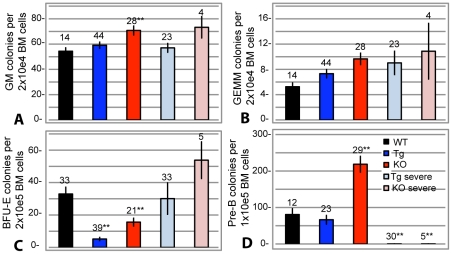
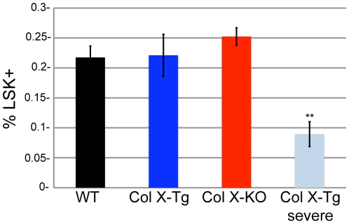
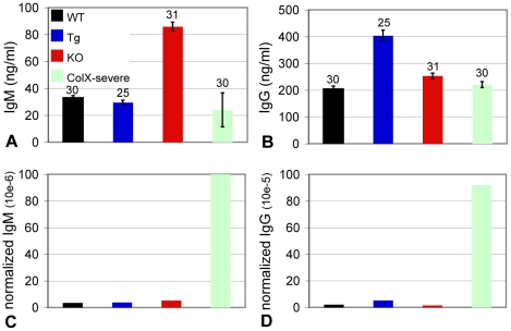
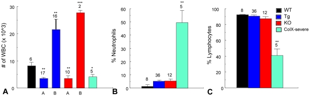
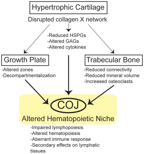
The link between endochondral skeletal development and hematopoiesis in the marrow was established in the collagen X transgenic (Tg) and null (KO) mice. Disrupted function of collagen X, a major hypertrophic cartilage matrix protein, resulted in skeletal and hematopoietic defects in endochondrally derived tissues. Manifestation of the disease phenotype was variable, ranging from perinatal lethality in a subset of mice, to altered lymphopoiesis and impaired immunity in the surviving mice. To exclude contribution of strain specific modifiers to this variable manifestation of the skeleto-hematopoietic phenotype, C57Bl/6 and DBA/2J collagen X congenic lines were established. Comparable disease manifestations confirmed that the skeleto-hematopoietic alterations are an inherent outcome of disrupted collagen X function. Further, colony forming cell assays, complete blood count analysis, serum antibody ELISA, and organ outgrowth studies established altered lymphopoiesis in all collagen X Tg and KO mice and implicated opportunistic infection as a contributor to the severe disease phenotype. These data support a model where endochondral ossification-specific collagen X contributes to the establishment of a hematopoietic niche at the chondro-osseous junction.


Conflict of interest statement

Competing Interests: The authors have declared that no competing interests exist.

Figures

Figure 1
Figure 1. Collagen X Tg and KO mice have an altered chondro-osseous junction.
Hematoxylin and eosin staining of longitudinal sections of tibia from (A) week-3 C57Bl/6 wild type (WT), (B) C57Bl/6 congenic collagen X transgenic (Tg), (C) null (KO), and perinatal lethal (D) Tg, and (E) KO. The chondro-osseous junction (COJ) is shown, including hypertrophic cartilage (HC), trabecular bone (TB), and marrow (M). Note diminished trabecular bony spicules in all collagen X Tg and KO mice, with the greatest reductions in (D) and (E), with a concomitant increase in erythrocytes in the marrow. Bar = 150 µm.
Figure 2
Figure 2. Collagen X Tg and KO mice have altered bone marrow cellularity and lymphocyte profiles.
(A) Geimsa staining of longitudinal sections of tibia from week-3 C57Bl/6 wild type (WT) and C57Bl/6 congenic collagen X transgenic (Tg), null (KO), and perinatal lethal (Tg severe, KO severe) mice showed mild hyperplasia in Tg and KO and aplasia with an influx of red blood cells in Tg severe and KO severe. (40X) Bar = 125 µm. (B) Temporal analysis by flow cytometry for B220+ B lymphocytes in bone marrow aspirates of outbred and congenic (C57Bl/6, B6 and DBA/2J, DBA) mice revealed reduced B220+ lymphocytes throughout life in Tg and KO, with an exception at week-3 in KO, in both the outbred and congenic strains. Note also acute B cell reduction in Tg severe and KO severe. (C) The total number of B220+ lymphocytes was calculated via flow cytometry and the trend of B lymphocyte reduction in the collagen X Tg and KO mice is maintained. Numbers above the standard error of the mean are the number of mice per group. (*p<0.05, **p<0.01)
Figure 3
Figure 3. Altered spleen architecture and B lymphocyte profile in the collagen X Tg and KO mice.
Giemsa and immunohistochemical staining of spleens from week-3 C57Bl/6 wild type (A, 2X; B, Hi mag) and congenic collagen X perinatal lethal mice (C, 2X; D, Hi mag) revealed diminished organs with altered tissue architecture in the perinatal lethal mice. Staining for red blood cells with TER119 antibody (upper halves of spleens on right of Giemsa stained sections) showed decreased zones of red pulp (RP) in perinatal lethal mouse spleen. Staining for B lymphocytes with B220 antibody (lower halves of spleens on right of Giemsa stained sections) revealed diffuse B220+ staining and a reduction of lymphatic node (N) size in perinatal lethal mouse spleen. Bar = 25 mm. (E) Temporal analysis by flow cytometry for B220+ B lymphocytes from of outbred and congenic (C57Bl/6, B6 and DBA/2J, DBA) wild type (WT) and collagen X transgenic (Tg), null (KO), and perinatal lethal (Tg severe, KO severe) mouse spleens revealed decreased B lymphocytes throughout life, with the exception of KO at weeks two and three, in both the outbred and congenic strains. (F) Number of B220+ B lymphocytes in congenic WT, collagen X Tg and KO mice. Note depletion of B lymphocytes in Tg severe and KO severe. Numbers above the standard error of the mean are the number of mice per group. (*p<0.05, **p<0.01)
Figure 4
Figure 4. Diminished number of immature thymocytes and altered thymic architecture in the collagen X perinatal lethal mice.
(A, B) Giemsa staining of thymuses from week-3 wild type (WT) and collagen X transgenic perinatal lethal (PL) mice reveals a reduction in size and depletion of the cortical thymocytes and region in the perinatal lethal mouse thymus, (2X) Bar = 775 µm and 20X respectively. (C) Flow cytometry analysis of week-3 C57Bl/6 wild type, congenic collagen X transgenic (Tg), null (KO) and perinatal lethal (Tg severe and KO severe) mouse thymocytes quantifies the percent double-positive, CD4+/CD8+, immature T lymphocytes (boxed). (D) Graphic representation of compiled flow cytometry data reveals decreased levels of CD4+/CD8+ immature T lymphocytes in perinatal lethal collagen X Tg and KO mice across all strains (outbred; OB, C57Bl/6; B6, and DBA/2J; DBA). n≥6 mice per group.
Figure 5
Figure 5. Colony forming cell assays confirm altered hematopoietic lineage commitment in the collagen X Tg and KO mice.
Colony forming assays using marrow aspirates from week-3 wild type (WT), collagen X transgenic (Tg), null (KO) and perinatal lethal (Tg severe and KO severe) mice. Cells cultured in: (A-B) complete Methocult media to enumerate granulocyte-macrophage (GM) and granulocyte-erythrocyte-monocyte-macrophage (GEMM) colonies; (C) erythropoietin supplemented Methocult media to enumerate pre-erythrocyte blast colonies (BFU-E); (D) interluekin-7 supplemented Methocult media to enumerate pre-B lymphocyte colonies (pre-B). Numbers above the standard error of the mean are number of samples. (*p<0.05, **p<0.001).
Figure 6
Figure 6. Number of LSK cells in collagen X moue bone marrow.
Quantification of lineage-, Sca-1+, cKit+ (LSK) cells in the bone marrow of week-3 wild type (WT), collagen X transgenic (Tg), collagen X null (KO) and the perinatal lethal collagen X Tg subset (Tg-severe) via flow cytometry. 10 mice per group. (**p<0.001)
Figure 7
Figure 7. Collagen X perinatal lethal mice have increased levels of serum antibodies.
Sera from week-3 wild type (WT), collagen X transgenic (Tg), null (KO) and perinatal lethal (Tg severe and KO severe; pooled) mice were assayed for IgM and IgG antibodies with ELISA. (A-B) Total IgM and IgG serum antibody measured. (C-D) Normalized levels of IgM and IgG antibodies calculated as (ng/ml of serum antibody)/(total number of live B220 splenocytes). Numbers above the standard error of the mean are number of mice per group.
Figure 8
Figure 8. Peripheral blood complete blood count (CBC) analysis implicates infections in collagen X perinatal lethal mice.
(A) White blood cell (WBC) counts from wild type (WT), collagen X transgenic (Tg), null (KO) and perinatal lethal (Tg severe and KO severe; pooled) reveal two groups of Tg and KO mice (A and B). (B) Percentage of neutrophils from peripheral blood reveals a thirteen-fold increase in the perinatal lethal subset. (C) Percentage of lymphocytes reveals a two-fold decrease in the perinatal lethal subset. Numbers above the standard deviations are number of mice per group (*p<0.05, **p<0.001).
Figure 9
Figure 9. All collagen X Tg and KO mice have skeleto-hematopoietic defects that directly arise from collagen X disruption in hypertrophic cartilage.
Disruption of collagen X function in hypertrophic cartilage of the growth plate results in the loss of a collagen X-containing pericellular network, leading to a reduction and altered compartmentalization of heparan sulfate proteoglycans (HSPGs) within the chondro-osseous junction (COJ) , as well as altered cytokine production by COJ cells ( and unpublished data). Resultant skeletal defects involve all EO-derived skeletal elements, and manifest as compressed growth plates , and reduction in the connectivity, mineral volume and overall amount of trabecular bone ( Fig. 1 and unpublished data). Collectively, these changes culminate in an altered COJ, which affects the differentiation of resident hematopoietic stem cells, thereby negatively effecting lymphopoiesis and immunity. Hematopoietic defects manifest as impaired lymphopoiesis in the marrow ( Fig. 2 ), with subtle effects on other blood cell lineages ( Fig. 5 ). Secondary hematopoietic/immune defects, arising as a consequence of an altered marrow environment, involve an impaired lymphocyte population in all lymphatic organs ( Figs. 3 , 4 ), and impaired immune response throughout life .

Similar articles

Cited by

References

    1. Chan D, Jacenko O. Phenotypic and biochemical consequences of collagen X mutations in mice and humans. Matrix Biol. 1998;17:169–184. - PubMed
    1. Lefebvre V, Smits P. Transcriptional control of chondrocyte fate and differentiation. Birth Defects Res Part C: Embryo Today: Reviews. 2005;75:200–212. - PubMed
    1. Aguila HL, Rowe DW. Skeletal development, bone remodeling, and hematopoiesis. Immunol Rev. 2005;208:7–18. - PubMed
    1. Jacenko O, Ito S, Olsen BR. Skeletal and hematopoietic defects in mice transgenic for collagen X. Ann N Y Acad Sci. 1996;785:278–280. - PubMed
    1. Jacenko O, Roberts DW, Campbell MR, McManus PM, Gress CJ, et al. Linking hematopoiesis to endochondral skeletogenesis through analysis of mice transgenic for collagen X. Am J Pathol. 2002;160:2019–2034. - PMC - PubMed

Publication types

Substances