. 2010 Mar 4;5(3):e9504.
doi: 10.1371/journal.pone.0009504.

A crucial role for Kupffer cell-derived galectin-9 in regulation of T cell immunity in hepatitis C infection

Affiliations

A crucial role for Kupffer cell-derived galectin-9 in regulation of T cell immunity in hepatitis C infection

John A Mengshol et al. PLoS One. .

Erratum in

  • PLoS One. 2010;5(3). doi: 10.1371/annotation/d15b793c-85c7-4529-bc80-aabcb088a8cf

Abstract

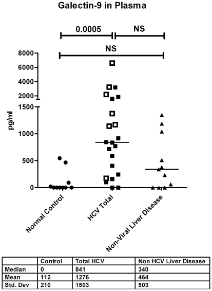
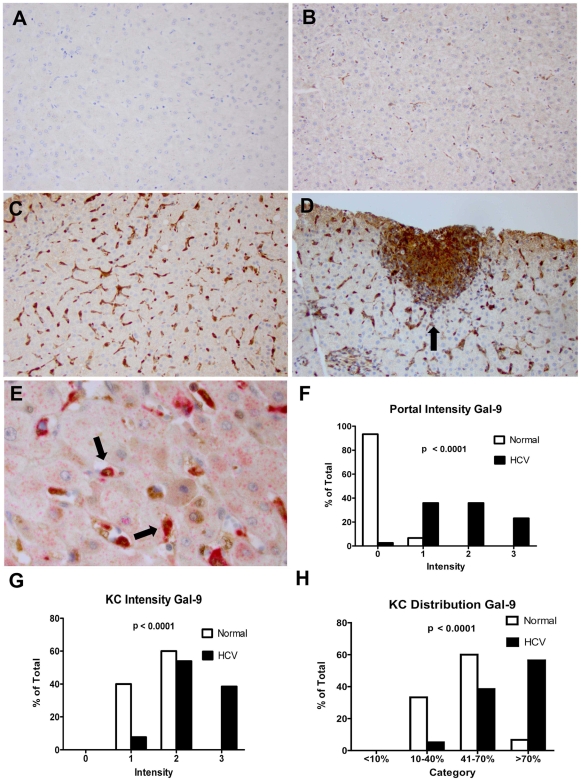
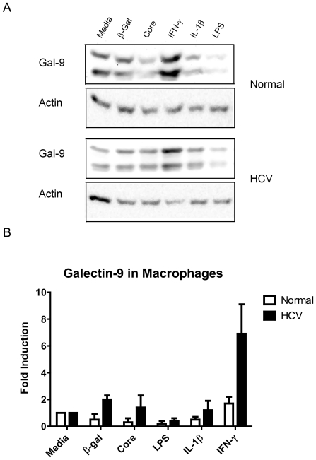
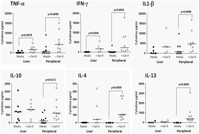
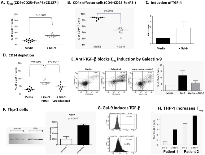
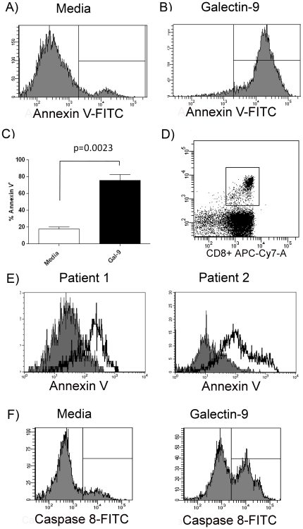
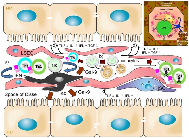
Approximately 200 million people throughout the world are infected with hepatitis C virus (HCV). One of the most striking features of HCV infection is its high propensity to establish persistence (approximately 70-80%) and progressive liver injury. Galectins are evolutionarily conserved glycan-binding proteins with diverse roles in innate and adaptive immune responses. Here, we demonstrate that galectin-9, the natural ligand for the T cell immunoglobulin domain and mucin domain protein 3 (Tim-3), circulates at very high levels in the serum and its hepatic expression (particularly on Kupffer cells) is significantly increased in patients with chronic HCV as compared to normal controls. Galectin-9 production from monocytes and macrophages is induced by IFN-gamma, which has been shown to be elevated in chronic HCV infection. In turn, galectin-9 induces pro-inflammatory cytokines in liver-derived and peripheral mononuclear cells; galectin-9 also induces anti-inflammatory cytokines from peripheral but not hepatic mononuclear cells. Galectin-9 results in expansion of CD4(+)CD25(+)FoxP3(+)CD127(low) regulatory T cells, contraction of CD4(+) effector T cells, and apoptosis of HCV-specific CTLs. In conclusion, galectin-9 production by Kupffer cells links the innate and adaptive immune response, providing a potential novel immunotherapeutic target in this common viral infection.


Conflict of interest statement

Competing Interests: Dr. Niki works for GalPharma and supplied antibodies for this study. This does not alter adherence to all the PLoS ONE policies on sharing data and materials, as detailed online.

Figures

Figure 1
Figure 1. Galectin-9 is elevated in the plasma of patients with chronic HCV and higher levels are seen in hepatocellular carcinoma (HCC).
Galectin-9 levels were analyzed in the plasma of 10 normal controls, 22 patients with HCV and 11 patients with non-viral causes of liver disease using a sandwich ELISA . Seven patients with HCV and HCC are denoted by open squares; they had significantly higher galectin-9 levels compared to HCV patients without HCC, p = 0.0334. Plasma from 11 patients with non-viral liver disease was analyzed. Three patients had alcoholic liver disease, 3 patients had Primary Biliary Cirrhosis, 3 patients had autoimmune hepatitis and 2 patients had non-alcoholic steatohepatitis. P- values were calculated using the two tailed Mann-Whitney test. NS denotes non-significant, p values>0.05.
Figure 2
Figure 2. HCV patients have increased Galectin-9 in Kupffer cells and periportal areas.
Immunohistochemistry of paraffin embedded samples (see Methods for details) was used to analyze 39 samples from HCV-infected patients and 15 normal subjects for galectin-9 staining. Staining for CD68 was used to identify Kupffer cells. The analyzing pathologist (M.S.) was blinded to patient identity and medical history. (A) Negative control with secondary antibody only, 20× magnification. (B) Galectin-9 staining in normal liver (brown) 20×; (C) Galectin-9 staining in a HCV patient 20×, (D) Periportal galectin-9 staining in HCV patient, 200×. (E) Co-localization of galectin-9 (brown) and CD68 (red) 600×. (F–H) Intensity of galectin-9 was scored from 0 to 3, and frequency was scored 0 (<10%), 1 (10–40%), 2 (40–70%), 3 (>70%). Distribution of scores by category expressed as a percent of the total patients comparing normal patients to patients with HCV. P-values calculated by the Chi-square test for trend.
Figure 3
Figure 3. Interferon gamma increases galectin-9 levels in macrophages.
CD14 positive cells from five normal controls and five HCV patients were bead selected from PBMCs and cultured for 48 hours with 25 ng/ml of MCSF to differentiate them into macrophages. Cells were stimulated for 48 hours with the stimuli shown at 2×10⁶/ml. IFN-γ was used at 25 ng/ml, HCV core and β-gal were used at 10 ug/ml, LPS was used at 100 ng/ml, and IL-1β was used at 10 ng/ml. Cell lysates were prepared and analyzed by Western Blot. (A) Western blot from one representative normal patient and one representative HCV patient to galectin-9 and β-Actin. (B) Fold induction was determined by densitometry using β-Actin as a loading control (n = 4 normal, and 3 HCV-positive patients).
Figure 4
Figure 4. Galectin-9 induces cytokine secretion from liver-derived and peripheral mononuclear cells.
Hepatic (n = 8) or peripheral (n = 8) mononuclear cells (2×10⁶/ml) were cultured in 96-well plates (200 ul/well) for 48 hours in media alone (RPMI+10% human serum) ± galectin-9 (5 ug/ml). After the culture period supernatants were collected and cytokine levels were measured using cytokine multiplex (Luminex™) technology as described in materials and methods. P-values were calculated using the Wilcoxan matched-pairs signed rank test.
Figure 5
Figure 5. Galectin-9 induces regulatory T cells (Tregs) via TGF-β.
Peripheral mononuclear cells (2×10⁶/ml) isolated from 5 chronic HCV patients and 6 normal controls were cultured for 5 days in media alone (RPMI+10% human serum) ± galectin-9 (5 ug/ml). After the culture period Treg levels were estimated using flow cytometry. Tregs were defined as CD4+CD25+FoxP3+CD127 T cells. Culture in the presence of galectin-9 induced Tregs (A) and a concomitant reduction in the CD4+CD25FoxP3 effector population was observed (B). (C) CD14+ monocytes were isolated from 3 chronic HCV patients and cultured in for 48 hours in media alone (RPMI+10% human serum) ± Galectin-9 (5 ug/ml). Galectin-9 increased the relative expression of TGF-β1 mRNA as analyzed by real time PCR. (D) Depletion of CD14+ cells from PBMC (8 normal patients) cultures treated with Galectin-9 attenuated the induction of Tregs at 5 days. (E) The addition of anti-TGF-β antibody at day 0 and day 2 of culture with PBMC and galectin-9 blocked induction of the Treg population. Representative flow plots are shown; the bar graphs are derived from three separate experiments. (F) THP-1 monocyte cell line constitutively expresses Galectin-9 which is upregulated by IFN-γ (25 ng/ml for 48 hours). Both the western blot and the densitometry analysis are shown, samples analyzed in triplicate. (G) Galectin-9 treatment induces TGF-β production from THP-1 cells. THP-1 cells were cultured with galectin-9 at 2.5 ug/ml for 24 hours then incubated with the latency associated peptide (LAP) then anti-LAP-PE to detect TGF-β. (H) IFN-γ treated THP-1 cells induce Treg from CD4+ T cells. Cells were cultured at a 1∶1 ratio for 5 days. Tregs were defined as CD4+CD25+FoxP3+CD127 T cells and graphed as a % of total CD4+ cells. P-values were calculated using the Wilcoxan matched-pairs signed rank test.
Figure 6
Figure 6. Galectin-9 induces apoptosis of HCV-specific CTLs through caspase-8 activation.
CD8+ T cells clones specific for NS3∶1406, NS3∶1436 and NS5∶2594 were incubated for 6 hours in media alone (A) or with 5 ug/ml of galectin-9 (B) followed by staining with Annexin V and 7-AAD. Representative histograms showing the percent of HCV-specific T cells staining with Annexin V following the indicated treatment. (C) Combined data for 5 CTL clones from different patients demonstrated an increase in Annexin V/7-AAD with galectin-9 treatment. P-values were calculated using the Wilcoxan matched-pairs signed rank test. (D) Plot showing NS3∶1436-specific CD8+ T cells by pentamer staining ex vivo. (E) Annexin V staining of NS3∶1436- specific CD8+ T cells from two HCV patients cultured for 6 hours with media (grey shading) or galectin-9 (solid line, no shading). (F). Galectin-9 induces T cell apoptosis through caspase-8 activation. Shown are PBMC from a HCV-positive patient treated for 1 hour with 5 ug/ml galectin-9 or media alone. A FITC-conjugated caspase-8 inhibitor that binds specifically to activated caspase-8 was added for the last hour of culture. Cells were then stained with anti-CD8 and 1436-pentamer and the percentage of HCV-specific T cells with activated caspase-8 was determined by flow cytometric analysis.
Figure 7
Figure 7. Paradigm for central role of galectin-9 in regulation of hepatitis C immunity.
Liver sinusoids are lined by a fenestrated layer of sinusoidal endothelial cells. The unique architecture of the liver allows interaction of antigen-presenting cells with T cells. a) Chronic HCV infection is characterized by hepatic infiltration of Th1, Tc1, NK and NKT cells that secrete IFN-γ which stimulates KCs to produce galectin-9. b) Galectin-9 (large brown arrows) expands CD4+CD25+FoxP3+CD127low regulatory T cells via TGF-β dependent mechanisms. c) HCV-specific CTLs expressing high levels of the Tim-3 receptor are engaged and become apoptotic. d) Galectin-9 also induces production of pro-inflammatory cytokines (TNF-α, IL-1β, IFN-γ) and pro-fibrotic cytokines (TGF-β) that can act on hepatocytes and hepatic stellate cells (HSC). The space of Disse contains the HSC. Hepatocytes (HC), liver sinusoidal endothelial cells (LSEC), Kupffer cells (KC), Th1 (CD4+ T cells), Tc1 (CD8+ T cells), natural killer (NK) and natural killer T (NKT) cells, hepatic stellate cells (HSC). Inset, the strategic location of KCs and the slow blood flow through the sinusoids allows contact with infiltrating lymphocytes (structural relationship of hepatic cells adapted from [18], [45]).

Similar articles

Cited by

References

    1. Shepard CW, Finelli L, Alter MJ. Global epidemiology of hepatitis C virus infection. Lancet Infect Dis. 2005;5:558–567. - PubMed
    1. Wang CC, Krantz E, Klarquist J, Krows M, McBride L, et al. Acute hepatitis C in a contemporary US cohort: modes of acquisition and factors influencing viral clearance. J Infect Dis. 2007;196:1474–1482. - PubMed
    1. Kim WR, Brown RS, Jr, Terrault NA, El-Serag H. Burden of liver disease in the United States: summary of a workshop. Hepatology. 2002;36:227–242. - PubMed
    1. Fried MW, Shiffman ML, Reddy KR, Smith C, Marinos G, et al. Peginterferon alfa-2a plus ribavirin for chronic hepatitis C virus infection. N Engl J Med. 2002;347:975–982. - PubMed
    1. Rabinovich GA, Toscano MA. Turning ‘sweet’ on immunity: galectin-glycan interactions in immune tolerance and inflammation. Nat Rev Immunol. 2009;9:338–352. - PubMed

Publication types

MeSH terms