. 2010 Mar 2;4(3):e618.
doi: 10.1371/journal.pntd.0000618.

Complete in vitro life cycle of Trypanosoma congolense: development of genetic tools

Affiliations

Complete in vitro life cycle of Trypanosoma congolense: development of genetic tools

Virginie Coustou et al. PLoS Negl Trop Dis. .

Abstract

Background: Animal African trypanosomosis, a disease mainly caused by the protozoan parasite Trypanosoma congolense, is a major constraint to livestock productivity and has a significant impact in the developing countries of Africa. RNA interference (RNAi) has been used to study gene function and identify drug and vaccine targets in a variety of organisms including trypanosomes. However, trypanosome RNAi studies have mainly been conducted in T. brucei, as a model for human infection, largely ignoring livestock parasites of economical importance such as T. congolense, which displays different pathogenesis profiles. The whole T. congolense life cycle can be completed in vitro, but this attractive model displayed important limitations: (i) genetic tools were currently limited to insect forms and production of modified infectious BSF through differentiation was never achieved, (ii) in vitro differentiation techniques lasted several months, (iii) absence of long-term bloodstream forms (BSF) in vitro culture prevented genomic analyses.

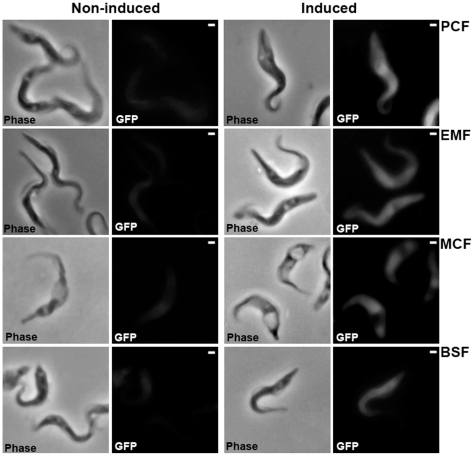
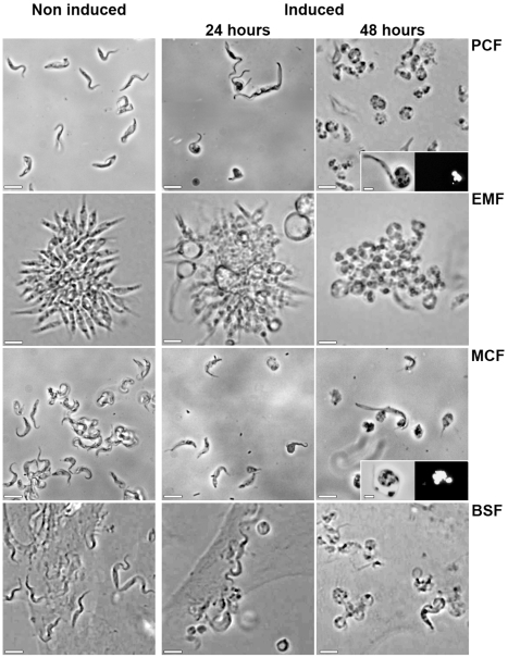
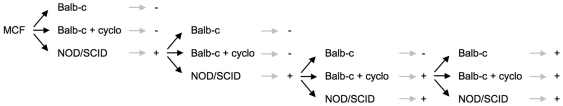
Methodology/principal findings: We optimized culture conditions for each developmental stage and secured the differentiation steps. Specifically, we devised a medium adapted for the strenuous development of stable long-term BSF culture. Using Amaxa nucleofection technology, we greatly improved the transfection rate of the insect form and designed an inducible transgene expression system using the IL3000 reference strain. We tested it by expression of reporter genes and through RNAi. Subsequently, we achieved the complete in vitro life cycle with dramatically shortened time requirements for various wild type and transgenic strains. Finally, we established the use of modified strains for experimental infections and underlined a host adaptation phase requirement.

Conclusions/significance: We devised an improved T. congolense model, which offers the opportunity to perform functional genomics analyses throughout the whole life cycle. It represents a very useful tool to understand pathogenesis mechanisms and to study potential therapeutic targets either in vitro or in vivo using a mouse model.


Conflict of interest statement

The authors have declared that no competing interests exist.

Figures

Figure 1
Figure 1. Inducible expression of EGFP through the T. congolense life cycle.
Microscope images of the in vitro cultivated IL3000:13–29 strain transfected with pLEW20c-EGFP vector in all the developmental stages. Non induced and tetracycline (1 µg/ml for 48 h) induced cells were fixed and visualized in phase contrast and by EGFP fluorescence (images were recorded with the same exposure time). Scale bars = 1 µm.
Figure 2
Figure 2. Inducible RNAi on tubulin genes through the T. congolense life cycle.
Phase contrast microscope images of the in vitro cultivated IL3000:13–29 strain transfected with p2T7Ti/αTUB vector in all the developmental stages. Non induced and tetracycline (1 µg/ml for 24 h and 48 h) induced cells are presented. PCF, EMF colonies, MCF (after DE52 purification) and BSF on BAE feeder cell layer were observed directly in the culture medium. In the insets, PCF and MCF were fixed and stained with 4,6-diamino-2-phenylindole (DAPI) before observation in phase contrast. Scale bars = 10 µm.
Figure 3
Figure 3. Morphological features and molecular markers analysis during metacyclogenesis.
A, Wild-type individual IL 3000 cells were labeled with monoclonal anti-PFR antibody during metacyclogenesis. For orientation the fixed cells were also stained with 4,6-diamino-2-phenylindole (DAPI) and visualized in phase contrast. Scale bar = 1 µm. B, Western blot analysis of stage specific markers during the metacyclogenesis. 106 cells of IL3000 were loaded per well. Tubulin was used as a loading control. Molecular weights are indicated on the blots side. iEMF: induced EMF in the absence of serum. Identical results were observed with the other T. congolense isolates and with transfected parasites.
Figure 4
Figure 4. Infectivity of TRUM183:13–29 cell-line through mice passages.
107 in vitro differentiated MCF or a volume corresponding to 107 parasites of infected mice blood were injected in Balb-c, Balb-c pretreated with cyclophosphamide or NOD/SCID mice. Development of parasitaemia was monitored by microscopic observation of mice blood every two days during 2 months. − means that no parasite could be observed during the time period of observation, + means that parasitaemia developed during the time period of observation.
Figure 5
Figure 5. Comparison of T. brucei and T. congolense in vitro growth.
A, T. brucei 427 BSF have been inoculated at 5.104 cell/ml in TcBSF-1 (Table 1) medium at 37°C in a humidified atmosphere containing 5% (vol/vol) CO2. B, T. congolense STIB910 BSF have been inoculated at 5.104 in presence of BAE (black curves) or at 2.105 cell/ml in absence of BAE (grey curves) in TcBSF-2 (Table 1) medium at 34°C in a humidified atmosphere containing 5% (vol/vol) CO2. Parasites were cultured in their specific medium for five days (——⧫) or in medium supplemented with red blood cell lysate (resuspension of red blood cells vol/vol in cold water and 100 fold diluted in medium) (——•), or with hemoglobin (100 µg/ml) (—formula image
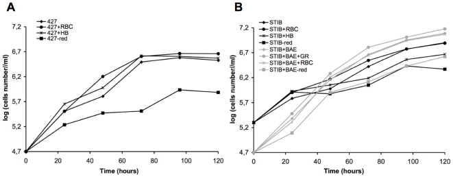
), or in absence of reducing agents (Bathocuproin, 2-mercaptoethanol and cystein were removed) (——▪). Cells were counted every 24 h.
Figure 6
Figure 6. In vitro culture system and genetic tools for T. congolense.
A, The different developmental stages cultured in vitro are represented as individual cells for PCF and MCF as adherent cells forming colonies for EMF and on BAE layer for BSF. Scale bar = 1 mm for PCF, MCF, BSF in infected mouse blood and the lower photo of BSF on BAE layer. PCF, EMF and BSF are dividing cells as represented by the rounded arrow. BSF differentiation can be achieved either by infection of mice and culture from blood or directly in vitro on BAE layer. B, Scheme of transgenic cell-line analysis through the cycle starting from PCF (top) or BSF (bottom) transfection. Linearized vectors used in transfection assays are represented by a line and two boxes, the black one represents the selection marker and the grey one represents the transgene (Tg).

Similar articles

Cited by

References

    1. Barrett MP, Boykin DW, Brun R, Tidwell RR. Human African trypanosomiasis: pharmacological re-engagement with a neglected disease. Br J Pharmacol. 2007;152:1155–1171. - PMC - PubMed
    1. Kappmeier K, Nevill EM, Bagnall RJ. Review of tsetse flies and trypanosomosis in South Africa. Onderstepoort J Vet Res. 1998;65:195–203. - PubMed
    1. Van den Bossche P. Some general aspects of the distribution and epidemiology of bovine trypanosomosis in southern Africa. Int J Parasitol. 2001;31:592–598. - PubMed
    1. LaCount DJ, Donelson JE. RNA interference in African trypanosomes. Protist. 2001;152:103–111. - PubMed
    1. Wirtz E, Clayton C. Inducible gene expression in trypanosomes mediated by a prokaryotic repressor. Science. 1995;268:1179–1183. - PubMed

Publication types

Substances