

The effect of superovulation on the contributions of individual blastomeres from 2-cell stage CF1 mouse embryos to the blastocyst
- PMID: 20209440
- PMCID: PMC9040203
- DOI: 10.1387/ijdb.092942mk
The effect of superovulation on the contributions of individual blastomeres from 2-cell stage CF1 mouse embryos to the blastocyst
Abstract
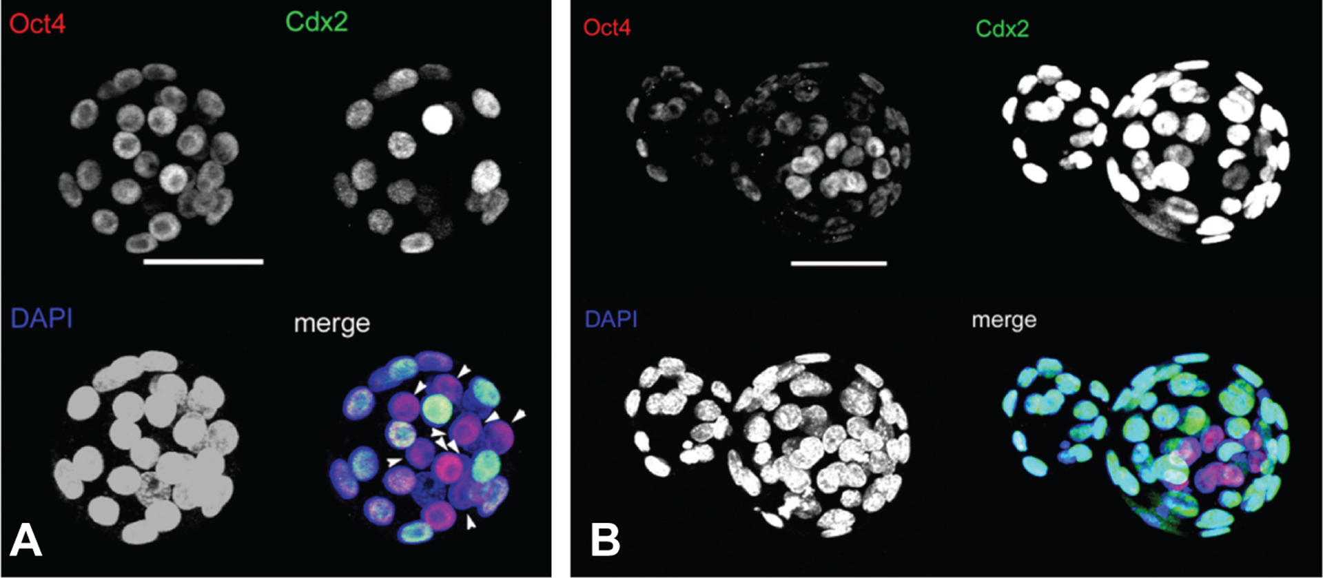
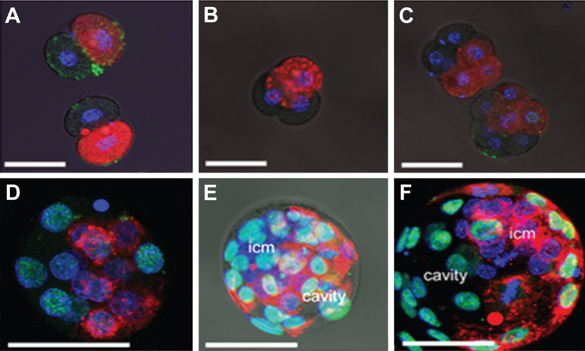
It remains controversial whether blastomeres of 2-cell stage mouse embryos show bias in their contribution to the blastocyst and whether there is any effect of superovulation. Two-cell stage embryos from CF1 mice were derived by either natural breeding (N) or superovulation (S) and cultured in vitro. At blastocyst, inner cell mass and trophectoderm were distinguished by Cdx2 and Oct4 immunostaining. A fluorescent dye (CM-Dil) was also used to tag individual blastomeres at the 2-cell stage, and the descendant cells identified by their red fluorescence. S and N embryos developed to blastocyst at the same rate and contained a similar number of cells. However, with S embryos, the descendants of the blastomere labeled with CM-DiI contributed predominantly to either the embryonic or abembryonic pole about 70% of the time, whereas most N embryos displayed random patterning, with no restriction to one or other of the poles. In S-embryos, but not N-embryos, the leading blastomere at second cleavage contributed preferentially to the embryonic pole of the blastocyst and the lagging blastomere to the abembryonic pole and hence mural trophectoderm. In addition, a tetrahedral rather than a flat morphology at the 4-cell stage of S-embryos was strongly biased to displaying the embryonic/abembryonic pattern at blastocyst. In contrast, S-embryos lacking a zona pellucida resembled N embryos in their patterning. In CF1 mice, superovulation has little effect on development to blastocyst, but enforces a greater degree of lineage restriction than natural breeding, most likely through constraints imposed by the zona pellucida.
Figures
Similar articles
-
Development of monozygotic twin mouse embryos from the time of blastomere separation at the two-cell stage to blastocyst.Biol Reprod. 2010 Jun;82(6):1237-47. doi: 10.1095/biolreprod.109.082982. Epub 2010 Feb 24. Biol Reprod. 2010. PMID: 20181620 Free PMC article.
-
Random Allocation of Blastomere Descendants to the Trophectoderm and ICM of the Bovine Blastocyst.Biol Reprod. 2016 Dec;95(6):123. doi: 10.1095/biolreprod.116.141200. Epub 2016 Oct 19. Biol Reprod. 2016. PMID: 27760750 Free PMC article.
-
Spatial alignment of the mouse blastocyst axis across the first cleavage plane is caused by mechanical constraint rather than developmental bias among blastomeres.Mol Reprod Dev. 2008 Jul;75(7):1143-53. doi: 10.1002/mrd.20856. Mol Reprod Dev. 2008. PMID: 18196554 Free PMC article.
-
Retrospective analysis: reproducibility of interblastomere differences of mRNA expression in 2-cell stage mouse embryos is remarkably poor due to combinatorial mechanisms of blastomere diversification.Mol Hum Reprod. 2018 Jul 1;24(7):388-400. doi: 10.1093/molehr/gay021. Mol Hum Reprod. 2018. PMID: 29746690
-
[Blastocyst Hatching in Humans].Ontogenez. 2017 Jan-Feb;48(1):8-20. Ontogenez. 2017. PMID: 30272915 Review. Russian.
Cited by
-
Effect of L-carnitine on in vitro developmental rate, the zona pellucida and hatching of blastocysts and their cell numbers in mouse embryos.Int J Reprod Biomed. 2016 Oct;14(10):649-656. Int J Reprod Biomed. 2016. PMID: 27921089 Free PMC article.
-
Totipotency segregates between the sister blastomeres of two-cell stage mouse embryos.Sci Rep. 2017 Aug 15;7(1):8299. doi: 10.1038/s41598-017-08266-6. Sci Rep. 2017. PMID: 28811525 Free PMC article.
-
Trophoblast stem cells.Biol Reprod. 2011 Mar;84(3):412-21. doi: 10.1095/biolreprod.110.088724. Epub 2010 Nov 24. Biol Reprod. 2011. PMID: 21106963 Free PMC article. Review.
-
Embryo cell allocation patterns are not altered by biopsy but can be linked with further development.Reproduction. 2017 Dec;154(6):807-814. doi: 10.1530/REP-17-0514. Epub 2017 Sep 29. Reproduction. 2017. PMID: 28971891 Free PMC article.
-
Development of monozygotic twin mouse embryos from the time of blastomere separation at the two-cell stage to blastocyst.Biol Reprod. 2010 Jun;82(6):1237-47. doi: 10.1095/biolreprod.109.082982. Epub 2010 Feb 24. Biol Reprod. 2010. PMID: 20181620 Free PMC article.
References
-
- ALARCON VB and MARIKAWA Y (2003). Deviation of the blastocyst axis from the first cleavage plane does not affect the quality of mouse postimplantation development. Biol Reprod 69: 1208–1212. - PubMed
-
- ALARCON VB and MARIKAWA Y (2005). Unbiased contribution of the first two blastomeres to mouse blastocyst development. Mol Reprod Dev 72: 354–361. - PubMed
-
- CHROSCICKA A, KOMOROWSKI S and MALESZEWSKI M (2004). Both blastomeres of the mouse 2-cell embryo contribute to the embryonic portion of the blastocyst. Mol Reprod Dev 68: 308–312. - PubMed
