. 2010 Aug 1;88(10):2103-17.
doi: 10.1002/jnr.22387.

Impaired neurogenesis is an early event in the etiology of familial Alzheimer's disease in transgenic mice

Affiliations

Impaired neurogenesis is an early event in the etiology of familial Alzheimer's disease in transgenic mice

Michael Demars et al. J Neurosci Res. .

Abstract

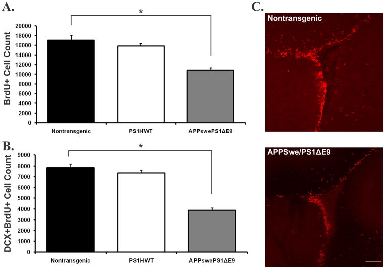
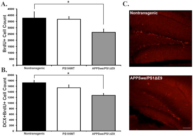
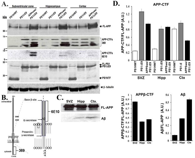
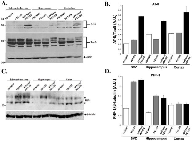
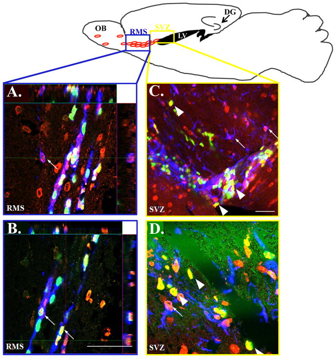
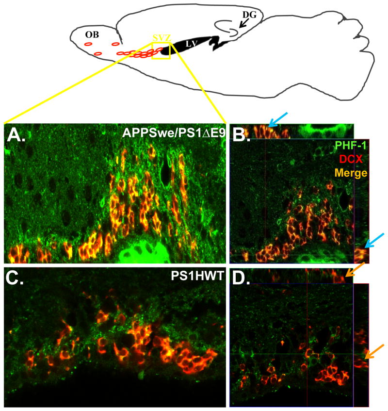
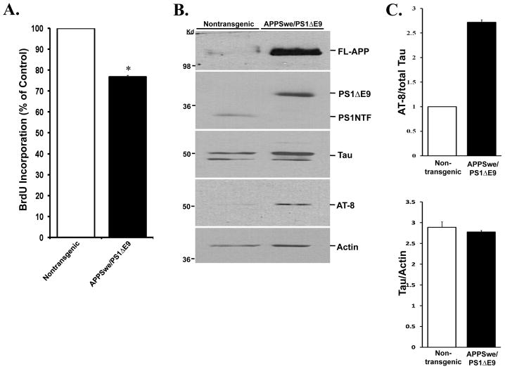
Formation of new neurons in the adult brain takes place in the subventricular zone and in the subgranule layer of the dentate gyrus throughout life. Neurogenesis is thought to play a role in hippocampus- and olfaction-dependent learning and memory. However, whether impairments in neurogenesis take place in learning and memory disorders, such as Alzheimer's disease, is yet to be established. Importantly, it remains to be elucidated whether neurogenic impairments play a role in the course of the disease or are the result of extensive neuropathology. We now report that transgenic mice harboring familial Alzheimer's disease-linked mutant APPswe/PS1DeltaE9 exhibit severe impairments in neurogenesis that are evident as early as 2 months of age. These mice exhibit a significant reduction in the proliferation of neural progenitor cells and their neuronal differentiation. Interestingly, levels of hyperphosphorylated tau, the cytotoxic precursor of the Alzheimer's disease hallmark neurofibrillary tangles, are particularly high in the neurogenic niches. Isolation of neural progenitor cells in culture reveals that APPswe/PS1DeltaE9-expressing neurospheres exhibit impaired proliferation and tau hyperphosphorylation compared with wildtype neurospheres isolated from nontransgenic littermates. This study suggests that impaired neurogenesis is an early critical event in the course of Alzheimer's disease that may underlie memory impairments, at least in part, and exacerbate neuronal vulnerability in the hippocampal formation and olfaction circuits. Furthermore, impaired neurogenesis is the result of both intrinsic pathology in neural progenitor cells and extrinsic neuropathology in the neurogenic niches. Finally, hyperphosphorylation of the microtubule-associated protein tau, a critical player in cell proliferation, neuronal maturation, and axonal transport, is a major contributor to impaired neurogenesis in Alzheimer's disease.


Figures

Figure 1
Figure 1. Reduced proliferation and neuronal differentiation is an early life primary neuropathological event in FAD-linked mice co-expressing APPswe and PS1ΔE9 mutations: the subventricular zone neurogenic microenvironemnt
(A) The number of BrdU immunoreactive cells is reduced by 36.09% in the subventricular zone of mice co-expressing APPswe/PS1ΔE9 mutations when compared with PS1HWT or non-transgenic littermates. (B) The number of BrdU immunoreactive cells that are co-labeled with the immature neuronal marker doublecortin (DCX) is dramatically reduced in APPswe/PS1ΔE9 mice when compared with PS1HWT or nontransgenic controls. (C) Representative images of BrdU immunoreactivity (red) in the subventricular zone of nontransgenic (top) and APPswe/PS1ΔE9 (bottom) mice (*p ≤ 0.005, students t-test). Scale bar= 100μm.
Figure 2
Figure 2. Reduced proliferation and neuronal differentiation is an early life primary neuropathological event in FAD-linked mice co-expressing APPswe and PS1ΔE9 mutations: the dentate gyrus neurogenic microenvironemnt
(A) The number of BrdU immunoreactive cells in the dentate gyrus of APPswe/PS1ΔE9 mice is greatly reduced when compared with PS1HWT or non-transgenic littermates. (B) As in the SVZ, the number of BrdU immunoreactive cells co-expressing doublecortin is also vastly reduced in the FAD-linked APPswe/PS1ΔE9 mice. (C) Representative images of BrdU immunoreactive cells in the dentate gyrus of nontransgenic (top) and APPswe/PS1ΔE9 (bottom) mice (*p ≤ 0.05, students t-test). Scale bar= 100μm.
Figure 3
Figure 3. Alterations in APP metabolites in the neurogenic microenvironments of APPswe/PS1ΔE9 mice
(A) Steady state levels of APP metabolites and PS1 in protein extract prepared from the SVZ, hippocampus and cortex of FAD-linked transgenic mice. FL-APP panel: Comparable expression levels of full-length APP in brain samples of PS1HWT and PS1ΔE9. Note over-expression of APP in transgenic mice harboring FAD-linked APPswe/PS1ΔE9. APP-CTF-369 panel: Increase in APP-CTFs in the cortex and neurogenic areas of APPswe/PS1ΔE9 due to transgene expression. APP-CTF-6E10 panel: Increase in APPswe-derived β-CTFs, in the SVZ of APPswe/PS1ΔE9 mice. PS1 panel: levels of transgenic PS1HWT N-terminal fragments (PS1NTF) and PS1ΔE9 are comparable in all brain areas. (B) Schematic presentation epitope binding site of 6E10 and 369 antibodies to APP. (C) Western blot analysis of soluble Aβ in protein extract prepared from the SVZ, hippocampus and cortex of APPswe/PS1ΔE9 mice revealing high levels of soluble Aβ in the cortex and hippocampus but virtually undetectable levels in the SVZ. Levels of full length APP (FL-APP) were comparable in the different regions. (D) Quantification of protein expression level of APP-CTFs relative to FL-APP (upper panel); APPβ-CTFs relative to FL-APP (bottom left panel); Aβ relative to FL-APP (bottom right panel). Error bars represent S.E.M.
Figure 4
Figure 4. Increased levels of phosphorylated tau in the neurogenic regions of APPswe/PS1ΔE9 mice
(A) Expression levels of total tau and phosphorylated tau in protein lysates of SVZ, hippocampus and cerebellum of PS1HWT, PS1ΔE9 and APPswe/PS1ΔE9 mice, as detected by Western blot analysis using tau-5 and AT-8 antibodies, respectively. A dramatic increase in tau phosphorylated at Ser-202/Thr-205 was detected by AT8 antibodies in all regions tested. (B) Quantification of the amount of AT8/actin relative to tau/actin. Error bars represent S.E.M. (C) Levels of phosphorylated tau in protein lysates of SVZ, hippocampus and cortex of PS1HWT, PS1ΔE9 and APPswe/PS1ΔE9 mice as detected by PHF-1 antibodies. Western blot analysis shows a marked increase in tau phosphorylated at Ser-396/Ser-404 in the neurogenic regions but not in the cortex. (D) Quantification of the relative amount of PHF-1 tau to β-tubulin in arbitrary units (A.U.).
Figure 5
Figure 5. Tau is expressed in neural stem cells, neural progenitor cells and migrating neuroblasts
TOP: Schematic presentation showing the neurogenic niches in a sagittal slice through the mouse brain: the region outlined by the blue box is representative of the area from which confocal RMS (A,B) images were taken and the yellow box represents the region corresponding to the SVZ images (C,D). Confocal imaging of immunolabeled brain sections of APPswe/PS1ΔE9 mice shows that tau co-localizes with doublecortin (small arrows; A,B), BrdU (A–D big arrows in C,D) and GFAP (small arrows; C,D). Tau, red (A–D); BrdU, green (A–D); doublecortin, blue (A,B); GFAP, blue (C,D). Scale bar= 50μm
Figure 6
Figure 6. PHF-1 is expressed in neuroblasts in the subventricular zone of APPswe/PS1ΔE9 mice
Upper Scheme: Schematic presentation of the area in the SVZ from which confocal images were taken. Left panel: PHF-1 expression is pronounced in DCX+ neuroblasts in the SVZ of APPswe/PS1ΔE9 (A) but hardly in PS1HWT (C) mice at 6 months of age. Right panel: High power orthogonal image showing strong co-localization of PHF-1 (green) and DCX (red) in the SVZ of APPswe/PS1ΔE9 (B) and no co-localization in PS1HWT mice (D). Blue arrows represent clear co-localization in the orthogonal view of APPSwe/PS1ΔE9 sections while the orange represent DCX+ cells in the PS1HWT SVZ that are clearly not PHF-1+. Scale bars= 50μm.
Figure 7
Figure 7. Neurospheres derived from the SVZ of APPSwe/PS1ΔE9 mice exhibit impaired proliferation and increased tau phosphorylation
(A) Proliferation assay examining BrdU incorporation in dissociated neurospheres shows a reduction in the proliferative capacity of neural progenitor cells derived from APPswe/PS1ΔE9 mice, compared to neural progenitor cells derived from nontransgenic littermates. (B) Western blot analysis of tau levels in protein extracts of neurospheres isolated from APPSwe/PS1ΔE9 and nontransgenic littermate mice. Total tau expression levels are comparable in neurospheres derived from APPSwe/PS1ΔE9 and nontransgenic mice compared to actin levels. However, a dramatic increase is detected in levels of phosphorylated tau in neurospehers derived APPSwe/PS1ΔE9 using AT-8 antibodies. Neurospheres isolated from APPSwe/PS1ΔE9 mice exhibit characteristic transgene expression pattern of APP (2–3 fold increase in FL-APP) and PS1 (lack of full length cleavage of PS1ΔE9). (C) Quantification of protein levels as detected in Western blot shows that the levels of AT8 are increased 3-fold relative to total tau (top) while tau levels are consistent when normalized to actin (bottom). Error bars represent S.E.M. (*p≤0.05, students t-test).

Similar articles

Cited by

References

    1. Aimone JB, Wiles J, Gage FH. Potential role for adult neurogenesis in the encoding of time in new memories. Nature neuroscience. 2006;9(6):723–727. - PubMed
    1. Albers MW, Tabert MH, Devanand DP. Olfactory dysfunction as a predictor of neurodegenerative disease. Curr Neurol Neurosci Rep. 2006;6(5):379–386. - PubMed
    1. Arriagada PV, Growdon JH, Hedley-Whyte ET, Hyman BT. Neurofibrillary tangles but not senile plaques parallel duration and severity of Alzheimer’s disease. Neurology. 1992;42(3 Pt 1):631–639. - PubMed
    1. Ashe KH. Learning and memory in transgenic mice modeling Alzheimer’s disease. Learning & memory (Cold Spring Harbor, NY. 2001;8(6):301–308. - PubMed
    1. Bacon AW, Bondi MW, Salmon DP, Murphy C. Very early changes in olfactory functioning due to Alzheimer’s disease and the role of apolipoprotein E in olfaction. Ann N Y Acad Sci. 1998;855:723–731. - PubMed

MeSH terms

LinkOut - more resources