

Review
doi: 10.1117/1.3285504.
Laser speckle contrast imaging in biomedical optics
Affiliations
- PMID: 20210435
- PMCID: PMC2816990
- DOI: 10.1117/1.3285504
Item in Clipboard
Review
Laser speckle contrast imaging in biomedical optics
J Biomed Opt.
2010 Jan-Feb.
Abstract
First introduced in the 1980s, laser speckle contrast imaging is a powerful tool for full-field imaging of blood flow. Recently laser speckle contrast imaging has gained increased attention, in part due to its rapid adoption for blood flow studies in the brain. We review the underlying physics of speckle contrast imaging and discuss recent developments to improve the quantitative accuracy of blood flow measures. We also review applications of laser speckle contrast imaging in neuroscience, dermatology and ophthalmology.
Figures
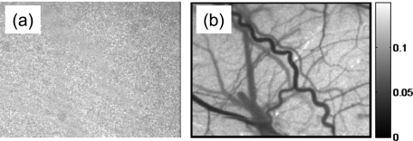
(a) Raw speckle image from the thin skull of a rat, showing a grainy pattern in which it is possible to discern some spatial variation in the speckle contrast, and (b) when the spatial speckle contrast is estimated from a 7×7 window of pixels, the blood vessels on the surface of the brain become apparent with high spatial resolution.
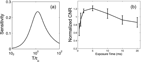
(a) Speckle contrast sensitivity to particle dynamics as given by camera integration time divided by decorrelation time and (b) the normalized contrast-to-noise ratio of changes in speckle contrast versus camera integration time due to changes in rodent cerebral blood flow. Reproduced from Ref. with permission.
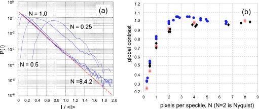
(a) Speckle intensity probability distribution function considering different numbers of pixels per speckle N and the distribution is an exponential for N=2. (b) The speckle contrast for a static medium equals its expected value of 1 when N=2. Reproduced from Ref. with permission.
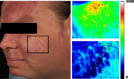
Illustration of LSCI for monitoring PWS treatment. Left: Photograph of patient with PWS in the area indicated by the rectangle. LSCI images were acquired immediately before (upper) and 15 minutes after (lower) laser therapy. Figure graciously provided by Bernard Choi.
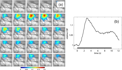
Imaging of stimulus induced changes in blood flow in the brain. (a) Sequence of images showing the percent changes in blood flow in response to 10 s of forepaw stimulation and (b) graph illustrating the percent change in blood flow over a 1.75×1.75-mm region of interest centered on the activation [see (a)]. Reproduced from Ref. with permission.
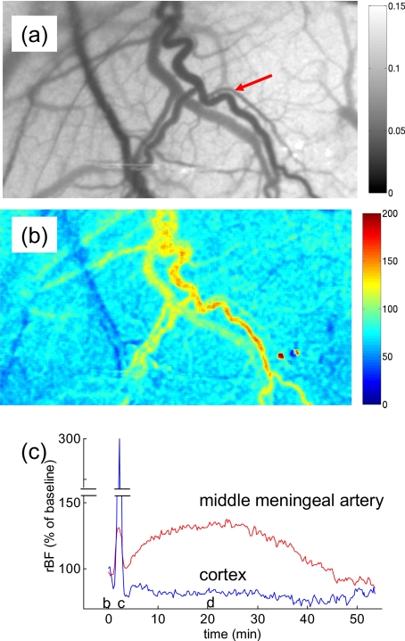
Blood flow images following cortical spreading depression: (a) the speckle contrast image reveals vasculature in the cortex and dura, the middle meningeal artery is indicated by the arrow; (b) relative blood flow 20 min after induction of cortical SD reveals elevated blood flow in the middle meningeal artery; and (c) time course of the changes in blood flow in the cortex and the middle meningeal artery demonstrating the long-lasting blood flow increase that is restricted to the dural vessel. Reproduced from Ref. with permission.
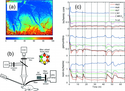
Application of LSCI to cerebral ischemia: (a) the spatial blood flow gradient following occlusion of an artery can be visualized using LSCI, where the middle cerebral artery was occluded just outside the top region of the image and the color map shows the relative blood flow, expressed as a percentage of preischemic flow; (b) LSCI and MSRI can be performed simultaneously to image multiple hemodynamic parameters; and (c) time courses of changes in oxyhemoglobin (HbO), deoxyhemoglobin (HbR), total hemoglobin (HbT), blood flow (CBF), oxygen consumption (CMRO2), and scattering during a stroke. The three graphs demonstrate the changes in each of these parameters in three spatial regions (ischemic core, penumbra, and nonischemic cortex). (b) and (c) reproduced from Ref. with permission.
References
-
- Cummins H. and Swinney H. L., “Light beating spectroscopy,” in Progress in Optics, Wolf E., Ed., Vol. VIII, p. 133, North Holland Publishing Co., Amsterdam: (1970).
-
- Riva C., Ross B., and Benedek G. B., “Laser Doppler measurements of blood flow in capillary tubes and retinal arteries,” Invest. Ophthalmol. INOPAO 11, 936–944 (1972). - PubMed
-
- Berne P. J. and Pecora R., Dynamic Light Scattering, Wiley, New York: (1976).
Publication types
MeSH terms
Grants and funding
LinkOut - more resources
Full Text Sources
Other Literature Sources
Medical
