

doi: 10.1117/1.3309455.
Detection of vesicant-induced upper airway mucosa damage in the hamster cheek pouch model using optical coherence tomography
Affiliations
- PMID: 20210463
- PMCID: PMC2839801
- DOI: 10.1117/1.3309455
Item in Clipboard
Detection of vesicant-induced upper airway mucosa damage in the hamster cheek pouch model using optical coherence tomography
J Biomed Opt.
2010 Jan-Feb.
Abstract
Hamster cheek pouches were exposed to 2-chloroethyl ethyl sulfide [CEES, half-mustard gas (HMG)] at a concentration of 0.4, 2.0, or 5.0 mg/ml for 1 or 5 min. Twenty-four hours post-HMG exposure, tissue damage was assessed by both stereomicrography and optical coherence tomography (OCT). Damage that was not visible on gross visual examination was apparent in the OCT images. Tissue changes were found to be dependent on both HMG concentration and exposure time. The submucosal and muscle layers of the cheek pouch tissue showed the greatest amount of structural alteration. Routine light microscope histology was performed to confirm the OCT observations.
Figures
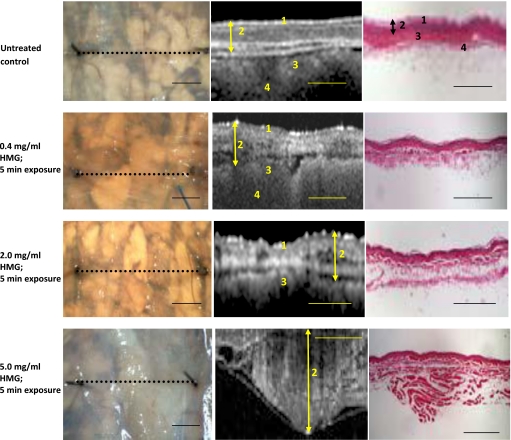
Stereomicrographs, OCT images, and corresponding histology of HMG treated hamster cheek pouch. Stereo micrographs (left column): Black dotted line=OCT imaging line. Arrows indicate visible blistering. OCT images (center column) and histological sections (right column): Double-ended arrow=maximum tissue depth of the mucosa. Total tissue thickness exceeds capturable OCT image size at the highest HMG concentration; surface epithelial layers are not visible in the OCT image. 1—Keratinized surface layer. 2—Flat stratified squamous epithelium. 3—Submucosa: dense fibrous connective tissue. 4—Longitudinal striated muscle. Calibration bars=1 mm.
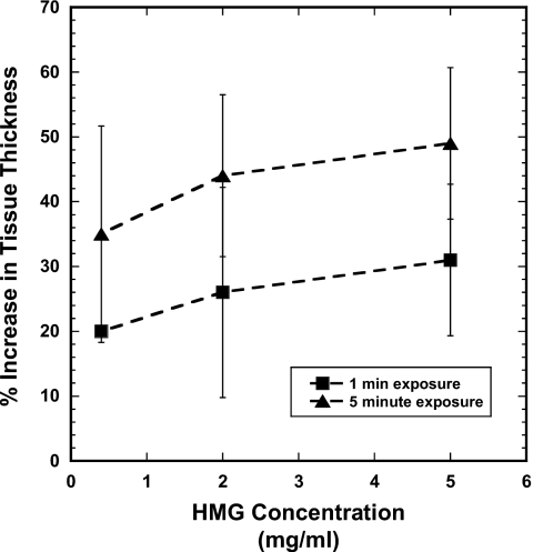
Tissue response to HMG exposure. Percent increase in total tissue thickness as a function of HMG concentration for 1-min and 5-min exposure times. Data points are mean±SEM.
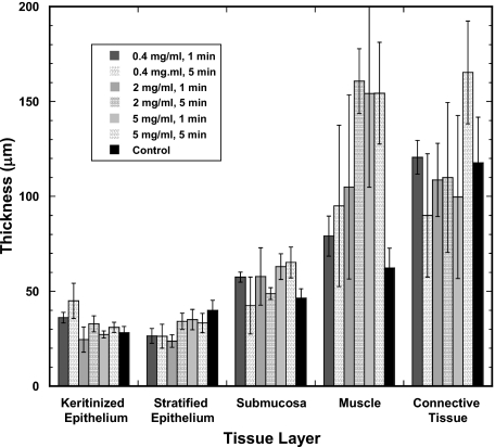
Tissue response by layer type. Thickness of each tissue layer of the hamster cheek pouch as measured on the OCT images following the application of HMG under various conditions. Values are mean±SEM.
Similar articles
-
Role of MAP kinases in regulating expression of antioxidants and inflammatory mediators in mouse keratinocytes following exposure to the half mustard, 2-chloroethyl ethyl sulfide.Toxicol Appl Pharmacol. 2010 Jun 15;245(3):352-60. doi: 10.1016/j.taap.2010.04.001. Epub 2010 Apr 9. Toxicol Appl Pharmacol. 2010. PMID: 20382172 Free PMC article.
-
Automated classification of optical coherence tomography images for the diagnosis of oral malignancy in the hamster cheek pouch.J Biomed Opt. 2014 Aug;19(8):086022. doi: 10.1117/1.JBO.19.8.086022. J Biomed Opt. 2014. PMID: 25162909 Free PMC article.
-
Optical coherence tomography of malignancy in hamster cheek pouches.J Biomed Opt. 2004 Sep-Oct;9(5):978-81. doi: 10.1117/1.1783897. J Biomed Opt. 2004. PMID: 15447019
-
The hamster cheek pouch model of carcinogenesis and chemoprevention.Adv Exp Med Biol. 1992;320:63-7. doi: 10.1007/978-1-4615-3468-6_9. Adv Exp Med Biol. 1992. PMID: 1279957 Review. No abstract available.
-
The hamster cheek pouch model for field cancerization studies.Periodontol 2000. 2015 Feb;67(1):292-311. doi: 10.1111/prd.12066. Periodontol 2000. 2015. PMID: 25494606 Review.
Cited by
-
Endoscopic Optical Coherence Tomography for Assessing Inhalation Airway Injury: A Technical Review.Otolaryngol (Sunnyvale). 2019;9(2):366. doi: 10.4172/2161-119X.1000366. Epub 2019 Apr 4. Otolaryngol (Sunnyvale). 2019. PMID: 31497378 Free PMC article.
References
-
- Compton J. A. F., Military Chemical and Biological Agents: Chemical and Toxicological Properties, pp. 5–17, Telford Press, Caldwell, NJ: (1998).
-
- Sohrabpour H., “Clinical manifestations of chemical agents on Iranian combatants during the Iran-Iraq conflict,” Arch. Belg. ARBEE6 291–297 (1989). - PubMed
-
- Wheeler G. P., “Studies related to the mechanisms of action of cytotoxic alkylating agents: a review,” Cancer Res. CNREA8 22, 651–688 (1962). - PubMed
-
- Dacre J. C. and Goldman M., “Toxicology and pharmacology of the chemical warfare agent sulfur mustard,” Pharmacol. Rev. ZZZZZZ 48, 289–326 (1996). - PubMed
