Detection of vesicant-induced upper airway mucosa damage in the hamster cheek pouch model using optical coherence tomography
- PMID: 20210463
- PMCID: PMC2839801
- DOI: 10.1117/1.3309455
Detection of vesicant-induced upper airway mucosa damage in the hamster cheek pouch model using optical coherence tomography
Abstract
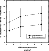
Hamster cheek pouches were exposed to 2-chloroethyl ethyl sulfide [CEES, half-mustard gas (HMG)] at a concentration of 0.4, 2.0, or 5.0 mg/ml for 1 or 5 min. Twenty-four hours post-HMG exposure, tissue damage was assessed by both stereomicrography and optical coherence tomography (OCT). Damage that was not visible on gross visual examination was apparent in the OCT images. Tissue changes were found to be dependent on both HMG concentration and exposure time. The submucosal and muscle layers of the cheek pouch tissue showed the greatest amount of structural alteration. Routine light microscope histology was performed to confirm the OCT observations.
Figures


References
-
- Compton J. A. F., Military Chemical and Biological Agents: Chemical and Toxicological Properties, pp. 5–17, Telford Press, Caldwell, NJ: (1998).
-
- Sohrabpour H., “Clinical manifestations of chemical agents on Iranian combatants during the Iran-Iraq conflict,” Arch. Belg. ARBEE6 291–297 (1989). - PubMed
-
- Wheeler G. P., “Studies related to the mechanisms of action of cytotoxic alkylating agents: a review,” Cancer Res. CNREA8 22, 651–688 (1962). - PubMed
-
- Dacre J. C. and Goldman M., “Toxicology and pharmacology of the chemical warfare agent sulfur mustard,” Pharmacol. Rev. ZZZZZZ 48, 289–326 (1996). - PubMed

