

Photoacoustic discrimination of vascular and pigmented lesions using classical and Bayesian methods
- PMID: 20210465
- PMCID: PMC2917464
- DOI: 10.1117/1.3316297
Photoacoustic discrimination of vascular and pigmented lesions using classical and Bayesian methods
Abstract
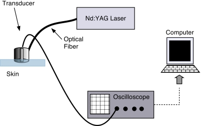
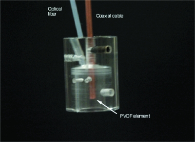
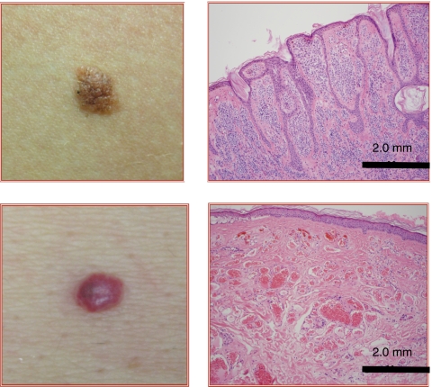
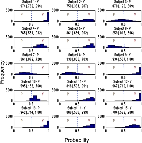
Discrimination of pigmented and vascular lesions in skin can be difficult due to factors such as size, subungual location, and the nature of lesions containing both melanin and vascularity. Misdiagnosis may lead to precancerous or cancerous lesions not receiving proper medical care. To aid in the rapid and accurate diagnosis of such pathologies, we develop a photoacoustic system to determine the nature of skin lesions in vivo. By irradiating skin with two laser wavelengths, 422 and 530 nm, we induce photoacoustic responses, and the relative response at these two wavelengths indicates whether the lesion is pigmented or vascular. This response is due to the distinct absorption spectrum of melanin and hemoglobin. In particular, pigmented lesions have ratios of photoacoustic amplitudes of approximately 1.4 to 1 at the two wavelengths, while vascular lesions have ratios of about 4.0 to 1. Furthermore, we consider two statistical methods for conducting classification of lesions: standard multivariate analysis classification techniques and a Bayesian-model-based approach. We study 15 human subjects with eight vascular and seven pigmented lesions. Using the classical method, we achieve a perfect classification rate, while the Bayesian approach has an error rate of 20%.
Figures

Similar articles
-
Towards noncontact skin melanoma selection by multispectral imaging analysis.J Biomed Opt. 2011 Jun;16(6):060502. doi: 10.1117/1.3584846. J Biomed Opt. 2011. PMID: 21721796
-
Reliability of computer image analysis of pigmented skin lesions of Australian adolescents.Cancer. 1996 Jul 15;78(2):252-7. doi: 10.1002/(SICI)1097-0142(19960715)78:2<252::AID-CNCR10>3.0.CO;2-V. Cancer. 1996. PMID: 8674000
-
Laser removal of pigmented and vascular lesions.J Cosmet Dermatol. 2006 Sep;5(3):204-9. doi: 10.1111/j.1473-2165.2006.00252.x. J Cosmet Dermatol. 2006. PMID: 17177741 Review.
-
Fractal and integer-dimensional geometric analysis of pigmented skin lesions.Am J Dermatopathol. 1995 Aug;17(4):374-8. doi: 10.1097/00000372-199508000-00012. Am J Dermatopathol. 1995. PMID: 8600802
-
Laser treatment of pigmented and vascular lesions in the office.Facial Plast Surg. 2004 Feb;20(1):63-9. doi: 10.1055/s-2004-822961. Facial Plast Surg. 2004. PMID: 15034816 Review.
Cited by
-
Melanin distribution from the dermal-epidermal junction to the stratum corneum: non-invasive in vivo assessment by fluorescence and Raman microspectroscopy.Sci Rep. 2020 Sep 1;10(1):14374. doi: 10.1038/s41598-020-71220-6. Sci Rep. 2020. PMID: 32873804 Free PMC article.
-
Non-invasive monitoring of ultrasound-stimulated microbubble radiation enhancement using photoacoustic imaging.Technol Cancer Res Treat. 2014 Oct;13(5):435-44. doi: 10.7785/tcrtexpress.2013.600266. Epub 2013 Aug 31. Technol Cancer Res Treat. 2014. PMID: 24000993 Free PMC article.
-
Predicting Metastasis in Melanoma by Enumerating Circulating Tumor Cells Using Photoacoustic Flow Cytometry.Lasers Surg Med. 2021 Apr;53(4):578-586. doi: 10.1002/lsm.23286. Epub 2020 Jun 18. Lasers Surg Med. 2021. PMID: 32557708 Free PMC article.
-
Evanescent Field Based Photoacoustics: Optical Property Evaluation at Surfaces.J Vis Exp. 2016 Jul 26;(113):54192. doi: 10.3791/54192. J Vis Exp. 2016. PMID: 27500652 Free PMC article.
-
State-of-the-Art Preclinical Photoacoustic Imaging in Oncology: Recent Advances in Cancer Theranostics.Contrast Media Mol Imaging. 2019 Apr 30;2019:5080267. doi: 10.1155/2019/5080267. eCollection 2019. Contrast Media Mol Imaging. 2019. PMID: 31182936 Free PMC article. Review.
References
Publication types
MeSH terms
Substances
Grants and funding
LinkOut - more resources
Full Text Sources
Other Literature Sources
Medical
