Review
. 2010 Aug 15;13(4):489-509.
doi: 10.1089/ars.2009.3021.

Redox control systems in the nucleus: mechanisms and functions

Affiliations
Review

Redox control systems in the nucleus: mechanisms and functions

Young-Mi Go et al. Antioxid Redox Signal. .

Abstract

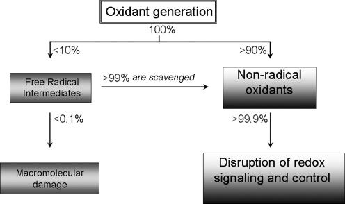
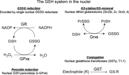
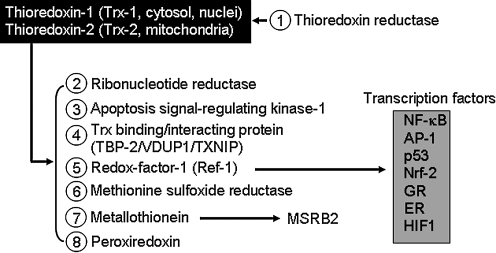
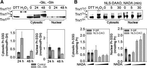
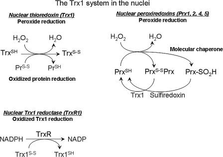
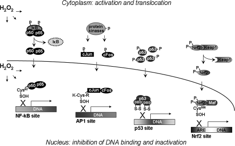
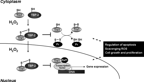
Proteins with oxidizable thiols are essential to many functions of cell nuclei, including transcription, chromatin stability, nuclear protein import and export, and DNA replication and repair. Control of the nuclear thiol-disulfide redox states involves both the elimination of oxidants to prevent oxidation and the reduction of oxidized thiols to restore function. These processes depend on the common thiol reductants, glutathione (GSH) and thioredoxin-1 (Trx1). Recent evidence shows that these systems are controlled independent of the cytoplasmic counterparts. In addition, the GSH and Trx1 couples are not in redox equilibrium, indicating that these reductants have nonredundant functions in their support of proteins involved in transcriptional regulation, nuclear protein trafficking, and DNA repair. Specific isoforms of glutathione peroxidases, glutathione S-transferases, and peroxiredoxins are enriched in nuclei, further supporting the interpretation that functions of the thiol-dependent systems in nuclei are at least quantitatively distinct, and probably also qualitatively distinct, from similar processes in the cytoplasm. Elucidation of the distinct nuclear functions and regulation of the thiol redox pathways in nuclei can be expected to improve understanding of nuclear processes and also to provide the basis for novel approaches to treat aging and disease processes associated with oxidative stress in the nuclei.


Figures

FIG. 1.
FIG. 1.
Mechanistic outcomes of oxidative stress include both macromolecular damage and disruption of redox signaling and control. Accumulating evidence indicates that both free radical (one-electron) and nonradical (two-electron) mechanisms contribute to damage from oxidative stress. Whereas superoxide anion radical and nitric oxide are free radicals, H2O2, lipid peroxides, quinones, epoxides, peroxynitrite, disulfides, and many other chemicals are nonradical oxidants. Based on model studies with xanthine oxidase, univalent reduction of O2 to superoxide anion radical is a minor fraction of the bivalent reduction to H2O2 (41), suggesting that two-electron oxidants predominate during oxidative stress. For an arbitrary rate of oxidant generation equal to 100%, perhaps 90% would go directly to two-electron oxidants, and only 10% would be one-electron oxidants. Because free radical–scavenging mechanisms are efficient and convert free radicals into nonradical oxidants, nonradical oxidants probably constitute >99.9% of all oxidants. Unlike reactive free radicals, which are nonspecific in their destruction of macromolecules, many of the nonradical oxidants selectively oxidize thiols. Many redox signaling and control mechanisms depend on critical cysteine residues. Consequently, disruption of redox signaling and control by nonradical oxidants may have a major role in oxidative stress as a mechanism of disease.
FIG. 2.
FIG. 2.
The GSH systems in the nuclei. Although systematic studies are limited, an extensive literature describes the GSH systems associated with nuclei. These include GSH, GSSG reductase (nuclear GSSG reductase), GSH peroxidase (n-Prx), glutaredoxins (Grx2b and Grx2c for human, Grx3 and Grx4 for yeast), and GST (GSTp and GST T1 are present in the nuclei). Many of these systems are identical or functionally equivalent to the cytoplasmic forms. However, the GSH systems exist under nonequilibrium redox conditions, so that function depends on abundance and distribution of individual redox components. In addition, changes in electron flow from NADPH or reduction of endogenously produced peroxide can change the steady-state redox potential and thereby control protein functions.
FIG. 3.
FIG. 3.
Thioredoxin-regulated cellular redox processes. (1) Thioredoxin (Trx) is reduced principally by thioredoxin reductases (TrxR). (2) Trx is an electron donor to support the activity of ribonucleotide reductase (RNR). (3) Trx binds to apoptosis signal-regulated kinase-1, inhibiting the kinase function. (4) Trx1 activity is inhibited by binding to Trx-binding protein-2 (TBP-2), also known as VDUP-1 and TXNIP. (5) Trx1 reduces redox factor-1 (Ref-1), a bifunctional protein with a redox activity and a separate DNA repair (APE-1, endonuclease) activity in nuclei; the redox activity maintains conserved Cys residues of transcription factors [NF-κB, AP-1, p53, Nrf2, glucocorticoid receptor (GR), estrogen receptor (ER), and HIF1] in the reduced form required for DNA binding. (6) Trx1 functions as a reductant for methionine sulfoxide reductases (MSRs). (7) Trx1 controls a pathway for MSRB2 reduction mediated by metallothionein. (8) Trx acts as a reductant for peroxiredoxin (Prx) activity to scavenge peroxides.
FIG. 4.
FIG. 4.
Quantification of nuclear redox state measured with Trx1 redox Western blotting and protein glutathionylation. Cells exposed to oxidative stress by depleting glucose (Glc) and glutamine (Gln) from culture (50) or by expressing nuclear-targeted d-amino acid oxidase (NLS-DAO) with variation of substrate amounts (N-acetyl-d-alanine, NADA) (56) were analyzed for cytoplasmic and nuclear redox-state measurements. (A) Nuclear Trx1 and GSH/GSSG were resistant to oxidation by Glc and Gln depletion, compared with cytoplasm. (B) Protein glutathionylation in nuclei was substantially increased by NLS-DAO expression, whereas no detectable change was seen in cytoplasmic protein glutathionylation or Trx1 oxidation in either cytoplasm or nuclei.
FIG. 5.
FIG. 5.
The thioredoxin-1 (Trx1) system in nuclei. The Trx system, including Trx1, thioredoxin reductase-1 (TrxR1), and Trx peroxidases (Prx1, Prx2, Prx4, and Prx5), is present in the nuclei. TrxR1 reduces oxidized Trx1 in the presence of NADPH. Trx1 functions to reduce peroxides and disulfide forms of oxidized proteins, such as Prx, which eliminates peroxides. Overoxidized Prx (Prx-SO2H) functions as a molecular chaperone and is reduced by sulfiredoxin.
FIG. 6.
FIG. 6.
A model for transcription-factor regulation in the cytoplasm and nuclei by oxidants. Cytoplasmic oxidants stimulate phosphorylation and translocation of transcription-factor proteins (NF-κB, AP-1, p53, and Nrf2) from the cytoplasm to nuclei to activate transcription. A nuclear oxidative mechanism oxidizes critical Cys residues in the DNA-binding region of the transcription factors and inhibits binding to DNA. This oxidative reaction inactivates transcriptional functions to turn off the system. Thus, the nuclear Trx1 and GSH systems have two functions in the nuclei: maintenance of the oxidant tone, which turns off the transcription factors, and repeated reduction of the critical Cys residues through Ref1 to maintain DNA binding.
FIG. 7.
FIG. 7.
Inhibition of Trx1 functions by TBP-2. Trx1-binding protein (TBP)-2 (also called VDUP1 or TXNIP) binds to Trx1 and inhibits Trx1 functions in reduction of protein disulfides. Oxidative stress stimulates this binding and also stimulates accumulation of the complex in nuclei. The Trx1/TBP complex accumulated in the nucleus by oxidants inhibits Trx1 interaction with Ref1. This process inhibits Trx1 function for Ref1 reduction, which in turn affects Ref1 activity to maintain Cys residues of transcription factors for DNA binding and activation for gene expression required for cell growth and proliferation.
FIG. 8.
FIG. 8.
Nuclear localization of Trx1 and glutathione is associated with cell proliferation. (A) Low cell-culture density, which is associated with oxidation of thiols and increased cell proliferation, affects the localization of Trx1 in cells. Each cell line (A549, Cos-1, HEK293) was plated in six-well plates containing glass coverslips at low and high density, incubated for 2 days, and assayed for Trx1 localization with immunofluorescence microscopy by using the Trx1 antibody. Data are reproduced from reference (177) with permission. (B) The nucleus/cytoplasm-to-GSH ratio, quantified with CMFDA fluorescence, shows higher levels in proliferating cells than in confluence, suggesting that a reduced nuclear environment is required for cell growth. Data are redrawn from reference (113).
FIG. 8.
FIG. 8.
Nuclear localization of Trx1 and glutathione is associated with cell proliferation. (A) Low cell-culture density, which is associated with oxidation of thiols and increased cell proliferation, affects the localization of Trx1 in cells. Each cell line (A549, Cos-1, HEK293) was plated in six-well plates containing glass coverslips at low and high density, incubated for 2 days, and assayed for Trx1 localization with immunofluorescence microscopy by using the Trx1 antibody. Data are reproduced from reference (177) with permission. (B) The nucleus/cytoplasm-to-GSH ratio, quantified with CMFDA fluorescence, shows higher levels in proliferating cells than in confluence, suggesting that a reduced nuclear environment is required for cell growth. Data are redrawn from reference (113).

Similar articles

Cited by

References

    1. Abate C. Patel L. Rauscher FJ., 3rd Curran T. Redox regulation of fos and jun DNA-binding activity in vitro. Science. 1990;249:1157–1161. - PubMed
    1. Ali-Osman F. Brunner JM. Kutluk TM. Hess K. Prognostic significance of glutathione S-transferase pi expression and subcellular localization in human gliomas. Clin Cancer Res. 1997;3:2253–2261. - PubMed
    1. Avval FZ. Holmgren A. Molecular mechanisms of thioredoxin and glutaredoxin as hydrogen donors for mammalian S phase ribonucleotide reductase. J Biol Chem. 2009;284:8233–8240. - PMC - PubMed
    1. Banmeyer I. Marchand C. Verhaeghe C. Vucic B. Rees JF. Knoops B. Overexpression of human peroxiredoxin 5 in subcellular compartments of Chinese hamster ovary cells: effects on cytotoxicity and DNA damage caused by peroxides. Free Radic Biol Med. 2004;36:65–77. - PubMed
    1. Bellomo G. Vairetti M. Stivala L. Mirabelli F. Richelmi P. Orrenius S. Demonstration of nuclear compartmentalization of glutathione in hepatocytes. Proc Natl Acad Sci U S A. 1992;89:4412–4416. - PMC - PubMed

Publication types

LinkOut - more resources