. 2010 Mar 8;3(1):6.
doi: 10.1186/1756-8935-3-6.

A paucity of heterochromatin at functional human neocentromeres

Affiliations

A paucity of heterochromatin at functional human neocentromeres

Alicia Alonso et al. Epigenetics Chromatin. .

Abstract

Background: Centromeres are responsible for the proper segregation of replicated chromatids during cell division. Neocentromeres are fully functional ectopic human centromeres that form on low-copy DNA sequences and permit analysis of centromere structure in relation to the underlying DNA sequence. Such structural analysis is not possible at endogenous centromeres because of the large amounts of repetitive alpha satellite DNA present.

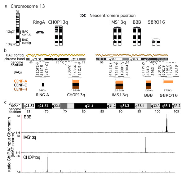
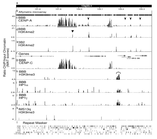
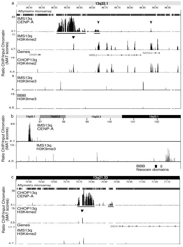
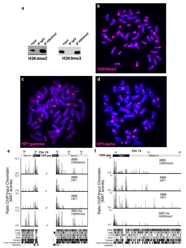
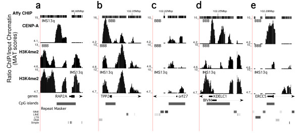
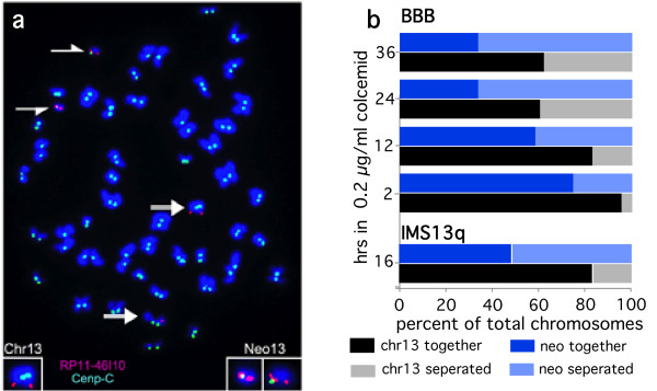
Results: High-resolution chromatin immunoprecipitation (ChIP) on CHIP (microarray) analysis of three independent neocentromeres from chromosome 13q revealed that each neocentromere contained approximately 100 kb of centromere protein (CENP)-A in a two-domain organization. Additional CENP-A domains were observed in the vicinity of neocentromeres, coinciding with CpG islands at the 5' end of genes. Analysis of histone H3 dimethylated at lysine 4 (H3K4me2) revealed small domains at each neocentromere. However, these domains of H3K4me2 were also found in the equivalent non-neocentric chromosomes. A surprisingly minimal (approximately 15 kb) heterochromatin domain was observed at one of the neocentromeres, which formed in an unusual transposon-free region distal to the CENP-A domains. Another neocentromere showed a distinct absence of nearby significant domains of heterochromatin. A subtle defect in centromere cohesion detected at these neocentromeres may be due to the paucity of heterochromatin domains.

Conclusions: This high-resolution mapping suggests that H3K4me2 does not seem sufficiently abundant to play a structural role at neocentromeres, as proposed for endogenous centromeres. Large domains of heterochromatin also do not appear necessary for centromere function. Thus, this study provides important insight into the structural requirements of human centromere function.


Figures

Figure 1
Figure 1
Chromatin immunoprecipitation (ChIP)-CHIP (microarray) analysis of chromosome 13q neocentromeres. (a) Ideograms of four inverted duplication 13q chromosomes, shown arched at the inversion breakpoint to indicate the duplicate regions homologous to (left) the normal chromosome 13. A ring chromosome derived from band 13q21 is also shown. The name of the cell line and the cytological position of each of the neocentromeres are indicated. The position of the 13q21 and 13q32 BAC microarrays are shown on the ideogram of the normal chromosome 13. (b) Expanded areas showing the 13q21 bacterial artificial chromosome (BAC) microarray (11 Mbp, 103 BACs)[12] and the 13q32 BAC microarray (14 Mbp, 126 BACs) [11]. The region that contained each neocentromere is expanded below, showing the BACs and their overlaps in each region. The positions of centromere protein (CENP)-A, -C and/or -H are shown when determined. The results showing colocalization of CENP-A, -C and -H on the IMS13q neocentromere are modified from a previous report [12]. (c) Affymetrix high density tiling array analysis of cell lines (CHOP13q, IMS13q and BBB), showing ~42 Mbp region encompassing the BAC microarrays and region between them on chromosome 13q21 to 13q33. The distinct and specific CENP-A domain identified for each neocentromere are shown. CENP-A chromatin immunoprecipitation (ChIP) model-based analysis of tiling-array (MAT) score: CHOP13q p < 10-10; IMS13q, P < 10-8; BBB, P < 10-8).
Figure 2
Figure 2
High-resolution analysis of chromatin domains at the BBB neocentromere. A 650 kb genomic region containing the neocentromere in cell line BBB, showing (a) the UCSC Hg18 genome coordinates from band 13q33.1 and (b) the coverage of the region on the Affymetrix tiling array, where gaps represent repetitive DNA not included on the array. (c) Results from chromatin immunoprecipitation (ChIP) with antibodies to centromere protein (CENP)-A from cell line BBB; model-based analysis of tiling-array (MAT) score. Horizontal lines indicate major and minor domains. Arrows indicate small domains that colocalize with the 5' end of the genes (see Figure 5). (d) ChIP with histone H3 dimethylated at lysine 4 (H3K4me2) antibody from cell line BBB;. Triangle indicates small domain within CENP-A domain. (e) ChIP with H3K4me2 antibody from fibroblast cell line 3B2, MAT P < 1.6 × 10⁻⁵, which serves as a non-neocentric control for BBB (f) The genes in the region. (g) ChIP with antibodies to CENP-C from cell line BBB. (h) ChIP with antibodies to H3K9me3 from cell line BBB. (i) ChIP with antibodies to heterochromatin protein (HP)1α from cell line BBB. (j) ChIP with antibodies to HP1γ from cell line BBB. (k) ChIP with antibodies to H3K9me3 from cell line IMS13q. (l) Repeat Masker tracks, where the 21.6 kb transposon-free region colocalizing with the H3K9me3 and HP1 domains can be seen. For domain coordinates and P values, see Table 1.
Figure 3
Figure 3
High-resolution analysis of chromatin domains at neocentromeres from IMS13q and CHOP13q cell lines. (a) A 650 kb genomic region surrounding the neocentromere in cell line IMS13q, showing the results for the chromatin immunoprecipitation (ChIP) analysis using antibodies to centromere protein (CENP)-A and H3K4me2. Arrows indicate small CENP-A domain colocalizing with the 5' end of genes (see Figure 5). Triangle indicates small histone H3 dimethylated at lysine 4 (H3K4me2) domain within the CENP-A domain. The H3K4me2 ChIP from CHOP13 serves as a control for IMS13q neocentromere region. H3K9me3 ChIP results for IMS13q and BBB are shown. (b) A 7 Mbp region containing the IMS13q CENP-A domain in band 13q32.1 and the H3K9me3 domain seen in this cell line in chromosome band 13q33.1. Note BBB CENP-A and H3K9me3 domain positions indicated (see Figure 2). (c) A 650 kb genomic region surrounding the neocentromere in cell line CHOP13q; ChIP for CENP-A and H3K4me2 is shown. The H3K4me2 ChIP from IMS13q serves as a control for CHOP13q neocentromere region. For domain coordinates and P values, see Table 1.
Figure 4
Figure 4
ChIP analysis at endogenous centromeres. (a) Western blot analysis of histone H3 dimethylated at lysine 4 (H3K4me2) and histone H3 methylated at lysine 9 (H3K9me) chromatin immunoprecipitation (ChIP). Lanes-Input chromatin, ChIP with rabbit IgG, and ChIP with antibody to (left) H3K4me2 or (right) H3K9me3. (b, c, d) DNA from the each ChIP experiment from the indicated antibody was labeled and used as a probe on metaphase spreads. H3K9me3, HP1α and HP1γ ChIP DNA hybridized to centromeric/pericentromeric regions. (e) An area of ~6 Mbp around the centromere gap of chromosome 10. (f) An area of ~2 Mbp at the centromere of chromosome 13. ChIP on CHIP (array) results for indicated antibodies and cell lines. All antibodies shown are enriched at the pericentromeric region. In chromosome 10, the pericentric heterochromatin is present over ~1 Mb at each side of the centromere gap. In chromosome 13, heterochromatin extends approximately 0.5 Mbp on the q arm. Note that genomic satellite DNA is not included on the Affymetrix CHIP. H3K9me3 (BBB) MAT score P < 2.5 × 10⁻⁵, HP1α (BBB) P < 5.6 × 10⁻³ and HP1γ (BBB) P < 9.6 × 10⁻³, H3K9me3 (IMS13q) P < 1.6 × 10⁻⁵. Repeat Masker tracks are shown below the graphs.
Figure 5
Figure 5
High-resolution analysis of centromere protein (CENP)-A and histone H3 dimethylated at lysine 4 (H3K4me2) at promoters in the vicinity of neocentromeres. Area of (a) ~4 kb at the promoter region of the RAP2A gene near the neocentromere in IMS13q (see Figure 3a); (b) ~4.8 kb at the promoter of the TPP2 gene in BBB (see Figure 2); (c) ~1500 bp at the c13orf gene in BBB; (d) ~4 kb at the KdelC1 and BIVM genes in BBB. (e) ~2200 bp at the ERCC5 gene in BBB. For each panel (a-e), the neocentric CENP-A domain is shown, and the H3K4me2 domains for both BBB and IMS13q lines. The transcription start site and CpG island for each gene are indicated. In general, the CENP-A is found in a distinct location from the H3K4me2 and appears to favor CpG islands.
Figure 6
Figure 6
Premature separation of neocentric metaphase chromosomes. (a) BBB cells treated with colcemid for ~24 hours show highly condensed chromosomes with separated arms but attached centromeres, as indicated by immunofluorescence with anti-centromere protein (CENP)-C (green). Fluorescent in situ hybridization (FISH) probe RP11-46I10 (red) from band 13q32 is found on the q arm of the two normal chromosomes 13 (full arrows) and on both ends of symmetric invdup13q neocentromere chromosome (see Fig 1). The neocentric chromosome has separated into its two replicated chromatids (split arrows), each of which contains a single kinetochore (green) which colocalizes with one of the RP11-46I10 FISH signals. (b) Quantification of separation of normal chromosome 13 (black and gray) and invdup13q neocentric (dark and light blue) chromosome at various times in colcemid. IMS13q at 16hours: 8/52 normal separated, 26/52 neocentric separated. BBB at 2 hours: 4/92 (4.5%) normal separated, 15/51 (30%) neocentric separated; at 12 hours: 17/103 (17%) normal separated, 26/57 (46%) neocentric separated; at 24 hours: 140/358 (39%) normal separated, 145/210 (70%) neocentric separated; at 36 hours: 41/108 (38%) normal separated, 39/56 (70%) neocentric separated.

References

    1. Black BE, Bassett EA. The histone variant CENP-A and centromere specification. Curr Opin Cell Biol. 2008;20:91–100. doi: 10.1016/j.ceb.2007.11.007. - DOI - PubMed
    1. Allshire RC, Karpen GH. Epigenetic regulation of centromeric chromatin: old dogs, new tricks? Nat Rev Genet. 2008;9:923–937. doi: 10.1038/nrg2466. - DOI - PMC - PubMed
    1. Foltz DR, Jansen LE, Black BE, Bailey AO, Yates JR, Cleveland DW. The human CENP-A centromeric nucleosome-associated complex. Nat Cell Biol. 2006;8:458–469. doi: 10.1038/ncb1397. - DOI - PubMed
    1. Bernard P, Allshire R. Centromeres become unstuck without heterochromatin. Trends Cell Biol. 2002;12:419–424. doi: 10.1016/S0962-8924(02)02344-9. - DOI - PubMed
    1. Sullivan BA, Karpen GH. Centromeric chromatin exhibits a histone modification pattern that is distinct from both euchromatin and heterochromatin. Nat Struct Mol Biol. 2004;11:1076–1083. doi: 10.1038/nsmb845. - DOI - PMC - PubMed

LinkOut - more resources