. 2010 Mar 8:7:17.
doi: 10.1186/1742-2094-7-17.

Reduction of beta-amyloid pathology by celastrol in a transgenic mouse model of Alzheimer's disease

Affiliations

Reduction of beta-amyloid pathology by celastrol in a transgenic mouse model of Alzheimer's disease

Daniel Paris et al. J Neuroinflammation. .

Abstract

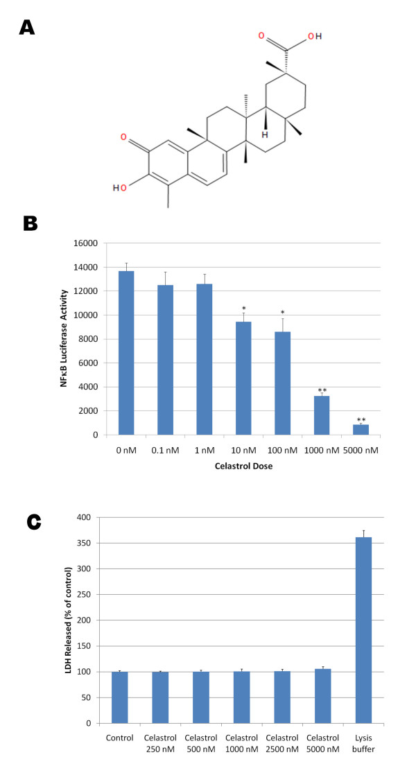
Background: Abeta deposits represent a neuropathological hallmark of Alzheimer's disease (AD). Both soluble and insoluble Abeta species are considered to be responsible for initiating the pathological cascade that eventually leads to AD. Therefore, the identification of therapeutic approaches that can lower Abeta production or accumulation remains a priority. NFkappaB has been shown to regulate BACE-1 expression level, the rate limiting enzyme responsible for the production of Abeta. We therefore explored whether the known NFkappaB inhibitor celastrol could represent a suitable compound for decreasing Abeta production and accumulation in vivo.

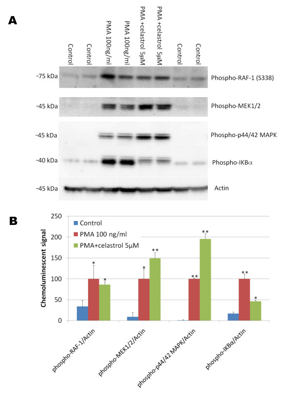
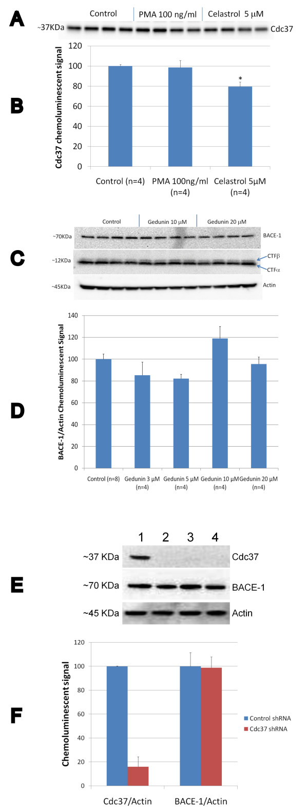
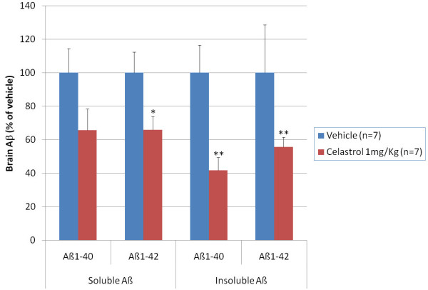
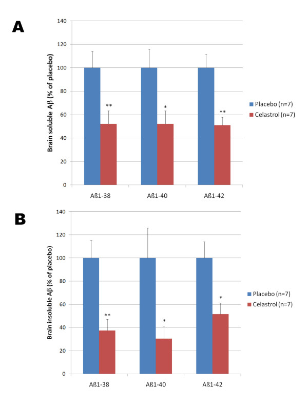
Methods: The effect of celastrol on amyloid precursor protein (APP) processing, Abeta production and NFkappaB activation was investigated by western blotting and ELISAs using a cell line overexpressing APP. The impact of celastrol on brain Abeta accumulation was tested in a transgenic mouse model of AD overexpressing the human APP695sw mutation and the presenilin-1 mutation M146L (Tg PS1/APPsw) by immunostaining and ELISAs. An acute treatment with celastrol was investigated by administering celastrol intraperitoneally at a dosage of 1 mg/Kg in 35 week-old Tg PS1/APPsw for 4 consecutive days. In addition, a chronic treatment (32 days) with celastrol was tested using a matrix-driven delivery pellet system implanted subcutaneously in 5 month-old Tg PS1/APPsw to ensure a continuous daily release of 2.5 mg/Kg of celastrol.

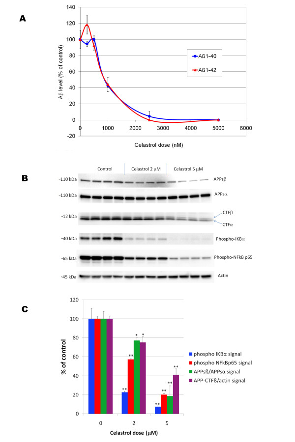
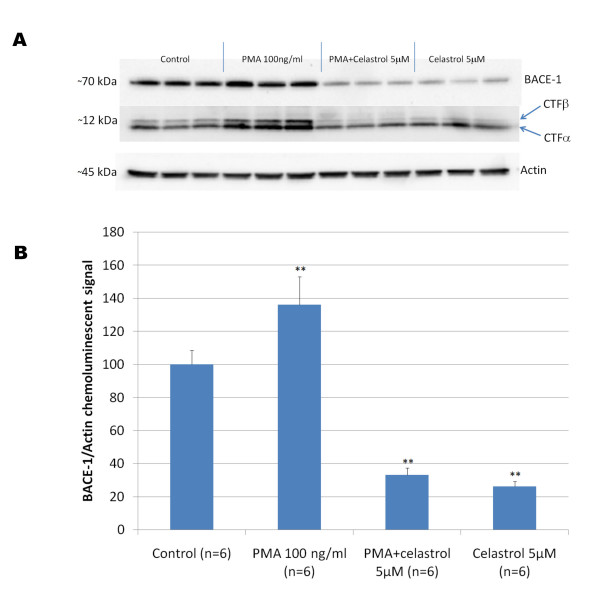
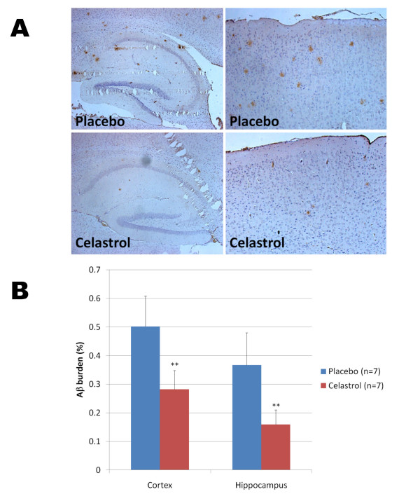
Results: In vitro, celastrol dose dependently prevented NFkappaB activation and inhibited BACE-1 expression. Celastrol potently inhibited Abeta1-40 and Abeta1-42 production by reducing the beta-cleavage of APP, leading to decreased levels of APP-CTFbeta and APPsbeta. In vivo, celastrol appeared to reduce the levels of both soluble and insoluble Abeta1-38, Abeta1-40 and Abeta1-42. In addition, a reduction in Abeta plaque burden and microglial activation was observed in the brains of Tg PS1/APPsw following a chronic administration of celastrol.

Conclusions: Overall our data suggest that celastrol is a potent Abeta lowering compound that acts as an indirect BACE-1 inhibitor possibly by regulating BACE-1 expression level via an NFkappaB dependent mechanism. Additional work is required to determine whether chronic administration of celastrol can be safely achieved with cognitive benefits in a transgenic mouse model of AD.


Figures

Figure 1
Figure 1
A) Chemical structure of celastrol, an active ingredient of the "Thunder of God" medication which has been used as an anti-inflammatory remedy for centuries in China. B) Dose dependent effect of celastrol on NFκB activation induced by TNFα in HEK293 cells stably expressing an NFκB luciferase reporter construct. Cells were co-treated with 20 pg/ml of TNFα and with a dose range of celastrol for 3 hours before measuring NFκB luciferase activity. ANOVA reveals a statistically significant main effect of celastrol dose on NFκB activation (P < 0.002). Post-hoc analysis show significant differences between NFκB activity in TNFα treated cells and cells co-treated with TNFα and celastrol for celastrol doses greater or equal to 10 nM (P < 0.005). C) The histogram represents the amounts of LDH released detected in the culture medium of HEK293 cells treated with a dose range of celastrol and with the MPER lysis buffer as a positive control (lysis buffer) to induce complete cellular lysis. ANOVA reveals no significant main effect of celastrol dose (P = 0.420) and a significant effect of the lysis buffer (P < 0.001) on LDH released.
Figure 2
Figure 2
Effect of celastrol on phorbol ester (PMA) induced phosphorylation of key members of the NFκB signaling pathway. HEK293 cells overexpressing APPsw were treated for 15 minutes with PMA in the presence of 0 μM and 5 μM of celastrol. A) Western blots reveal that celastrol did not inhibit Raf-1 phosphorylation, MEK1/2 phosphorylation or p44/42 MAPK phosphorylation induced by PMA but prevented IKBα phosphorylation induced by PMA suggesting that celastrol is inhibiting IKK activity. B) Histogram representing the quantification of Raf-1 phosphorylation/actin, MEK1/2 phosphorylation/actin, phospho p44/42 MAPK/actin and IKBα phosphorylation/actin chemoluminescent signal. ANOVA shows a significant main effect of PMA on the phosphorylation of these different proteins (P < 0.05). Post-hoc comparisons reveal significant increase in phosphorylation after PMA treatment for Raf-1 (P < 0.04), MEK1/2 (P < 0.01), p44/42 MAPK (P < 0.001) and IKBα (P < 0.001) compared to the control conditions. Celastrol does not appear to prevent PMA induced Raf-1 phosphorylation (P = 0.619) but leads to a significant stimulation of MEK1/2 and p44/42 MAPK compared to PMA treatment alone (P < 0.02) and significantly inhibited IKBα phosphorylation compared to PMA treatment alone (P < 0.001).
Figure 3
Figure 3
A) Effect of celastrol on Aβ production in 7 W CHO cells overexpressing wild-type human APP. Human Aβ was measured by ELISAs in the culture media surrounding the cells following 24 hours of treatment with different doses of celastrol. Dose response curves for both Aβ1-40 and Aβ1-42 were established revealing an IC50 of approximately 900 nM for celastrol. ANOVA reveals a significant main effect of celastrol on Aβ 1-40 (P < 0.001) and Aβ 1-42 production (P < 0.015). Post-hoc comparisons show statistically significant effects of celastrol at 1000 nM, 25000 nM and 5000 nM for both Aβ 1-40 and Aβ 1-42 (P < 0.001). B) Effect of celastrol on APP processing and NFκB in CHO cells overexpressing wild-type human APP. Celastrol dose dependently inhibited APPsβ secretion, APP-CTFβ level as well as NFκB p65 and IKBα phosphorylation. C) Histogram representing the quantification of APPsβ/APPsα, APP-CTFβ/actin as well as phospho-NFκB p65/actin and phospho-IKBα/actin chemoluminescent signals. ANOVA reveals a significant main effect of celastrol on APPsβ secretion, APP-CTFβ level, phospho-NFκB p65 and phospho-IKBα levels (P < 0.001). Post-hoc comparisons show a statistical significance for celastrol at 2 and 5 μM for all the parameters studied (P < 0.05) showing that celastrol dose dependently inhibits the β-cleavage of APP while suppressing NFκB activity. (* P < 0.05; ** P < 0.001).
Figure 4
Figure 4
A) Representative western-blot depicting the effect of celastrol on BACE-1 expression in HEK293 cells overexpressing APPsw. Celastrol inhibited basal BACE-1 expression as well as the stimulation of BACE-1 expression induced by a 24 hours treatment with PMA. APP-CTFβ level is reduced when BACE-1 expression is inhibited by celastrol and APP-CTFβ level is increased when BACE-1 expression is stimulated by PMA. B) Histogram representing the quantification of BACE-1 expression in response to celastrol and PMA treatments. ANOVA reveals a significant main effect of PMA (P < 0.002) and of celastrol (P < 0.001) on BACE-1 expression. Post-hoc comparisons shows that PMA significantly stimulates BACE-1 level (P < 0.002) whereas celastrol significantly reduces BACE-1 expression (P < 0.001) compared to control condition and opposes BACE-1 stimulation induced by PMA (P < 0.001). (** P < 0.002).
Figure 5
Figure 5
Effect of celastrol and PMA on the formation of the HSP90-cdc37 complex in HEK293 APPsw cells. A) Western-blot depicting the amount of cdc37 recovered in the HSP90 immunoprecipitate following treatment with celastrol and PMA. B) Histogram representing the quantification of cdc37 in the HSP90 immunoprecipitate. No significant effect of PMA was observed but a significant effect of celastrol was detected (P < 0.005) showing a slight reduction in the amount of cdc37 present in the HSP90 immunoprecipitates. C) Western-blots showing the effects of the HSP90 inhibitor gedunin on BACE-1 expression and APP processing in HEK293 APPsw cells. D) Histogram representing the quantification of BACE-1/Actin chemoluminescent signal showing that the gedunin treatment does not affect BACE-1 expression in HEK293 APPsw cells (ANOVA reveals no significant main effect of gedunin on BACE-1 level (P = 0.572)). E) Representative western-blot showing the level of cdc37 and BACE-1 expression in HEK293 APPsw cells that were knock-down for cdc37 using a shRNA approach for 3 different clones. 1) non silencing scrambled shRNA; 2) cdc37 shRNA clone 51G9; 3) cdc37 shRNA clone 97H1; 4) cdc37 shRNA clone 95C4. F) Histogram representing the quantification of cdc37 and BACE-1 expression for 9 different clones of HEK293 APPsw cells stably transfected with a silencing cdc37 shRNA vector (cdc37 shRNA) and 4 different clones of HEK293 APPsw stably transfected with a non silencing scrambled shRNA vector (control shRNA). Statistically significant inhibition of cdc37 expression (P < 0.001) and no effect on BACE-1 expression (P = 0.947) was observed in HEK293 APPsw cells knock-down for cdc37.
Figure 6
Figure 6
Acute effects of celastrol on brain Aβ levels in 35 week-old Tg PS1/APPSw. Mice were treated for 4 days with an intraperitoneal injection of celastrol (1 mg/Kg of body weight) or the vehicle only (placebo). Multivariate analysis reveals a significant main effect of celastrol on brain soluble and insoluble Aβ levels (P < 0.05). Statistically significant differences were observed for soluble Aβ 1-42 values (P < 0.03) between vehicle and celastrol treated mice and for insoluble Aβ 1-40 (P < 0.001) and Aβ 1-42 values (P < 0.001) between vehicle and celastrol treated mice. (* P < 0.05; ** P < 0.001).
Figure 7
Figure 7
Chronic effects of celastrol on brain Aβ levels. Five month-old Tg PS1/APPsw mice were implanted subcutaneously with biodegradable placebo pellets and celastrol pellets ensuring a constant release of celastrol at a rate of 2.5 mg/Kg of body weight/day. After 32 days of treatment, brain Aβ levels were analyzed by electrochemoluminescence. A) Histogram representing the level of soluble Aβ 1-38, Aβ 1-40 and Aβ 1-42 quantified in the brain of placebo and celastrol treated Tg PS1/APPsw mice. B) Histogram representing the level of insoluble Aβ 1-38, Aβ 1-40 and Aβ 1-42 quantified in the brain of placebo and celastrol treated Tg PS1/APPsw mice. Multivariate analysis reveals a statistical significant differences between celastrol and placebo treated mice for the levels of brain soluble Aβ 1-38 (P < 0.005), Aβ 1-40 (P < 0.03), Aβ 1-42 (P < 0.007) and brain insoluble Aβ 1-38 (P < 0.004), Aβ 1-40 (P < 0.03), Aβ 1-42 (P < 0.02). (* P < 0.05); ** P < 0.01).
Figure 8
Figure 8
Chronic effects of celastrol on Aβ plaque burden in Tg PS1/APPsw mice. A) Representative photomicrographs (200× magnification) showing the extent of Aβ plaque burden detected by 4G8 immunostaining in the cortex (right panel) and hippocampus (left panel) of Tg PS1/APPsw mice (6 month-old) treated with biodegradable pellets of placebo or celastrol for a period of 32 days. B) Histogram representing the quantification of Aβ burden by image analysis in the cortex and hippocampus of Tg PS1/APPsw mice treated with placebo and celastrol pellets. Multivariate analysis reveals a statistically significant effect of celastrol treatment on Aβ plaque burden for the cortex (P < 0.002) and hippocampus (P < 0.004). (**P < 0.005).
Figure 9
Figure 9
Chronic effects of celastrol on microgliosis in Tg PS1/APPsw mice. A) Representative photomicrographs (taken with a 20× and 60× objective providing a respective magnification of 200× and 600× respectively) depicting the presence of CD45 reactive microglia around Aβ deposits in Tg PS1/APPsw treated with a placebo and celastrol. B) Histogram representing the burden of activated microglia (CD45 positive) in the cortex of Tg PS1/APPsw mice treated with placebo and celastrol pellets. Statistically significant difference in microgliosis burden (P < 0.03) was observed between placebo and celastrol treated mice. (* P < 0.05).

References

    1. Selkoe DJ. Soluble oligomers of the amyloid beta-protein impair synaptic plasticity and behavior. Behav Brain Res. 2008;192(1):106–13. doi: 10.1016/j.bbr.2008.02.016. - DOI - PMC - PubMed
    1. Vassar R, Bennett BD, Babu-Khan S, Kahn S, Mendiaz EA, Denis P, Teplow DB, Ross S, Amarante P, Loeloff R, Luo Y, Fisher S, Fuller J, Edenson S, Lile J, Jarosinski MA, Biere AL, Curran E, Burgess T, Louis JC, Collins F, Treanor J, Rogers G, Citron M. Beta-secretase cleavage of Alzheimer's amyloid precursor protein by the transmembrane aspartic protease BACE. Science. 1999;286(5440):735–41. doi: 10.1126/science.286.5440.735. - DOI - PubMed
    1. Sinha S, Anderson JP, Barbour R, Basi GS, Caccavello R, Davis D, Doan M, Dovey HF, Frigon N, Hong J, Jacobson-Croak K, Jewett N, Keim P, Knops J, Lieberburg I, Power M, Tan H, Tatsuno G, Tung J, Schenk D, Seubert P, Suomensaari SM, Wang S, Walker D, Zhao J, McConlogue L, John V. Purification and cloning of amyloid precursor protein beta-secretase from human brain. Nature. 1999;402(6761):537–40. doi: 10.1038/990114. - DOI - PubMed
    1. Yan R, Bienkowski MJ, Shuck ME, Miao H, Tory MC, Pauley AM, Brashier JR, Stratman NC, Mathews WR, Buhl AE, Carter DB, Tomasselli AG, Parodi LA, Heinrikson RL, Gurney ME. Membrane-anchored aspartyl protease with Alzheimer's disease beta-secretase activity. Nature. 1999;402(6761):533–7. doi: 10.1038/990107. - DOI - PubMed
    1. Cole SL, Vassar R. The role of amyloid precursor protein processing by BACE1, the beta-secretase, in Alzheimer disease pathophysiology. J Biol Chem. 2008;283(44):29621–5. doi: 10.1074/jbc.R800015200. - DOI - PMC - PubMed

Publication types

MeSH terms