. 2010 Mar 8:10:84.
doi: 10.1186/1471-2407-10-84.

Enhanced antiproliferative and apoptotic response to combined treatment of gamma-tocotrienol with erlotinib or gefitinib in mammary tumor cells

Affiliations

Enhanced antiproliferative and apoptotic response to combined treatment of gamma-tocotrienol with erlotinib or gefitinib in mammary tumor cells

Sunitha V Bachawal et al. BMC Cancer. .

Abstract

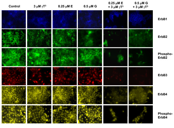
Background: Aberrant ErbB receptor signaling is associated with various types of malignancies. gamma-Tocotrienol is a member of the vitamin E family of compounds that displays potent anticancer activity that is associated with suppression in ErbB receptor phosphorylation and mitogenic signaling. Erlotinib and gefitinib are tyrosine kinase inhibitors that block ErbB1 receptor activation, whereas trastuzumab is a monoclonal antibody that has been designed to specifically inhibit ErbB2 receptor activation. However, the clinical effectiveness of these agents have been disappointing because of cooperation between different ErbB family members that can rescue cancer cells from agents directed against a single ErbB receptor subtype. It was hypothesized that targeting multiple ErbB receptor subtypes with combined treatment of gamma-tocotrienol and ErbB receptor inhibitors would provide greater anticancer effects than monotherapy targeting only a single ErbB receptor subtype.

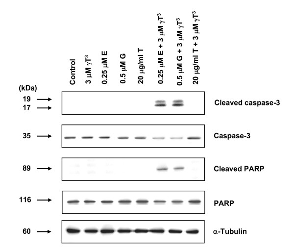
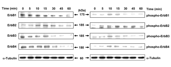
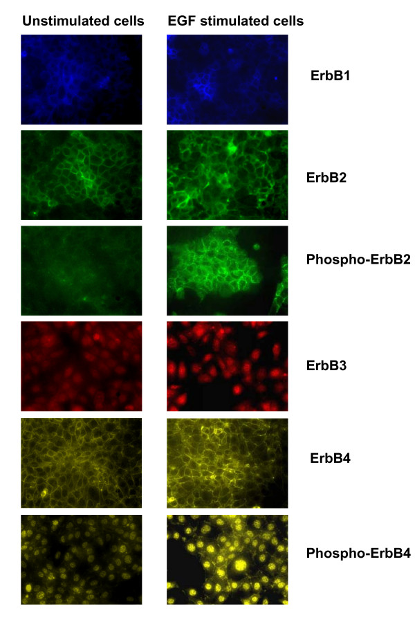
Methods: Highly malignant mouse +SA mammary epithelial cells were maintained in culture on serum-free defined media containing 10 ng/ml EGF as a mitogen. Cell viability wase determined by MTT assay, whereas Western blot and immunofluorescent staining was used to determine treatment effects on ErbB receptor subtype level and activation. Treatment-induced apoptosis was determined using annexin V staining and Western blot analysis of cleaved caspase-3 and PARP levels.

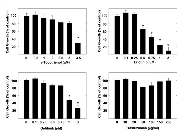
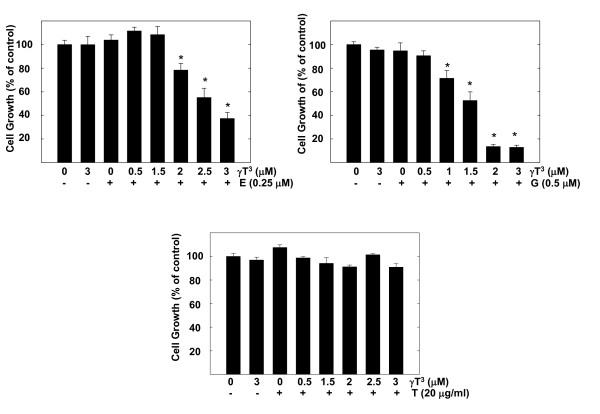
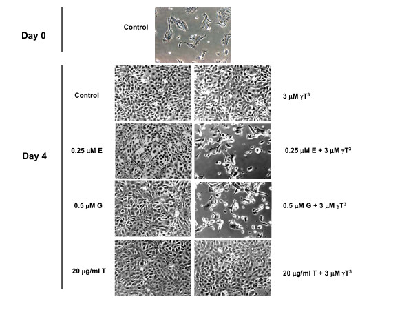
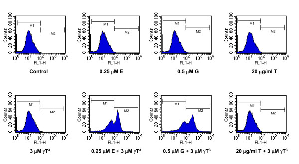
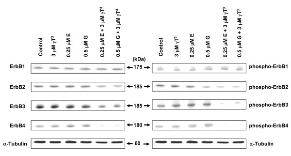
Results: Treatment with 3.5 microM gamma-tocotrienol, 0.5 microM erlotinib or 1.0 microM gefitinib alone, significantly inhibited +SA tumor cell growth. Combined treatment with subeffective doses of erlotinib (0.25 microM) or gefitinib (0.5 microM) with subeffective doses of gamma-tocotrienol (0.5-3.0 microM) significantly inhibited the growth and induced apoptosis in a dose-responsive manner. Trastuzumab treatment alone or in combination had no effect on +SA cell growth and viability. Combined treatment of gamma-tocotrienol with erlotinib or gefitinib also cause a large decrease in ErbB3, ErbB4, and to a lesser extent ErbB2 receptor levels, and EGF-dependent ErbB2-4 tyrosine phosphorylation (activation), but had no effect on ErbB1 receptor levels or activation.

Conclusion: Combination treatment of gamma-tocotrienol with specific ErbB receptor inhibitors is more effective in reducing mammary tumor cell growth and viability than high dose monotherapy, suggesting that targeting multiple ErbB receptors with combination therapy may significantly improve the therapeutic response in breast cancer patients.


Figures

Figure 1
Figure 1
Antiproliferative effects of γ-tocotrienol, erlotinib, gefitinib, and trastuzumab on neoplastic +SA mammary epithelial cell growth. Cells were plated at a density of 5 × 10⁴ cells per well (6 replicates/group) in 24-well plates and exposed to their respective treatments for a 4-day period. Viable cell number was determined by MTT colorimetric assay and indicated as percentage of control. Each bar represents the mean ± SEM in each treatment group and is representative of three independent experiments. *P < 0.05 as compared to the vehicle treated controls.
Figure 2
Figure 2
Effects of various subeffective doses of γ-tocotrienol (γT3) when given alone or in combination with subeffective dose of erlotinib (E), gefitinib (G), or trastuzumab (T) on neoplastic +SA mammary epithelial cell growth. Cells were plated at a density of 5 × 10⁴ cells per well (6 replicates/group) in 24-well plates and exposed to respective control and treatment serum-free defined media for a 4-day period. Viable cell number was determined by MTT colorimetric assay and indicated as percentage of control. Each bar represents the mean ± SEM in each treatment group and is representative of three independent experiments. *P < 0.05 as compared to the vehicle treated controls.
Figure 3
Figure 3
Photomicrographs of neoplastic +SA mammary epithelial cells in culture treated with a subeffective dose of γ-tocotrienol (γT3) alone or in combination with a subeffective dose of erlotinib (E), gefitinib (G), or trastuzumab (T). Cells were plated at a density of 5 × 10⁴ cells (Day 0) per well (6 replicates/group) in 24-well plates and exposed to respective control and treatment serum-free defined media for a 4-day period. Magnification is 100×.
Figure 4
Figure 4
Western blot analysis of γ-tocotrienol (γ T3), erlotinib (E), gefitinib (G), or trastuzumab (T) treatment effects given alone and in combination on the relative levels of cleaved caspase-3, caspase-3, cleaved PARP and PARP in neoplastic +SA mammary epithelial cells after a 4-day incubation period. +SA mammary tumor cells were plated at a density of 1 × 10⁶ cells/100 mm culture dish and incubated with control or treatment defined media over a period of 4-days. Afterwards, whole cell lysates (30 μg) from different treatment groups were prepared for subsequent separation by PAGE followed by Western blot analysis. Each Western blot is a representative image of data obtained for experiments that were repeated at least three times.
Figure 5
Figure 5
Effects of combined γ-tocotrienol (γT3), erlotinib (E), gefitinib (G), or trastuzumab (T) treatment on positive annexin V staining in +SA cells after a 4-day incubation period. +SA mammary tumor cells were plated at a density of 1 × 10⁶ cells/100-mm culture dish and incubated with control or treatment media over a 4-day period. Afterwards, cells were isolated with trypsin, and 1 × 10⁶ cells/group in triplicate were stained with annexin V, and then analyzed by flow cytometry. Viable cells display very low annexin V staining and appear in the marker 1 (M1) region, whereas cells undergoing apoptosis show very high annexin V staining and appear in the marker 2 (M2) region of the histogram.
Figure 6
Figure 6
Total ErbB and phosphorylated ErbB (activation) receptor levels in neoplastic +SA mammary epithelial cells. +SA mammary tumor cells were initially plated a density of 1 × 10⁶ cells/100 mm culture dish. Afterwards, cells were incubated overnight in mitogen-free media. Cells in all treatment groups were then stimulated with 100 ng/ml EGF for 0-60 min. Following treatment exposure, whole cell lysates were prepared and then subjected to PAGE followed by Western blot analysis for total and phosphorylated (active) ErbB receptor levels. Each Western blot is a representative image of data obtained for experiments that were repeated at least three times.
Figure 7
Figure 7
Immunofluorescent staining of ErbB1-4 receptors, phospho-ErbB2 and phospho-ErbB4 receptors. +SA mammary tumor cells were initially plated at a density of 2 × 10⁵ cells/well in 6-well plates (3 replicates/group) and allowed to attach to the plates followed by incubation in mitogen-free media overnight. Afterwards, cells were stimulated with 100 ng/ml EGF for 10 min. The cells were fixed with methanol and incubated with primary antibody directed against specific ErbB receptors, and then incubated with rhodamine conjugated secondary antibody, as described in the Methods section. Magnification is 200×.
Figure 8
Figure 8
Western blot analysis of total and phosphorylated ErbB receptor levels treated with γ-tocotrienol (γT3), erlotinib (E) or gefitinib (G) either alone or in combination. +SA mammary tumor cells were plated at a density of 1 × 10⁶ cells/100-mm culture dish and treated with control or treatment serum-free defined media containing EGF as mitogen. After a 4-day incubation period, whole cell lysates were subjected to PAGE followed by Western blot analysis for total and phosphorylated (active) ErbB receptor levels. Each Western blot is a representative example of data obtained for experiments that were repeated at least three times.
Figure 9
Figure 9
Immunofluorescent staining of ErbB1-4 receptors, phospho-ErbB2 and phospho-ErbB4 receptors in +SA cells after treatment with, γ-tocotrienol (γT3), erlotinib (E), gefitinib (G), alone or in combination. +SA mammary tumor cells were plated at a density of 2 × 10⁵ cells per well in 6 well plate (3 replicates/group) and incubated with control or treatment media for 4 days. The cells were then fixed with methanol and incubated with primary antibody directed against specific ErbB receptors followed by rhodamine conjugated secondary antibody, as described in the Methods section. Magnification is 200×.

Similar articles

Cited by

References

    1. McIntyre BS, Briski KP, Tirmenstein MA, Fariss MW, Gapor A, Sylvester PW. Antiproliferative and apoptotic effects of tocopherols and tocotrienols on normal mouse mammary epithelial cells. Lipids. 2000;35:171–180. doi: 10.1007/BF02664767. - DOI - PubMed
    1. McIntyre BS, Briski KP, Gapor A, Sylvester PW. Antiproliferative and apoptotic effects of tocopherols and tocotrienols on preneoplastic and neoplastic mouse mammary epithelial cells. Proc Soc Exp Biol Med. 2000;224:292–301. doi: 10.1046/j.1525-1373.2000.22434.x. - DOI - PubMed
    1. Sylvester PW, Shah SJ. Mechanisms mediating the antiproliferative and apoptotic effects of vitamin E in mammary cancer cells. Front Biosci. 2005;10:699–709. doi: 10.2741/1565. - DOI - PubMed
    1. Schneider C. Chemistry and biology of vitamin E. Mol Nutr Food Res. 2005;49:7–30. doi: 10.1002/mnfr.200400049. - DOI - PubMed
    1. Samant GV, Sylvester PW. γ-Tocotrienol inhibits ErbB3-dependent PI3K/Akt mitogenic signalling in neoplastic mammary epithelial cells. Cell Prolif. 2006;39:563–574. doi: 10.1111/j.1365-2184.2006.00412.x. - DOI - PMC - PubMed

Publication types

MeSH terms