. 2010 Jun 1;48(11):1548-58.
doi: 10.1016/j.freeradbiomed.2010.02.038. Epub 2010 Mar 6.

Nitric oxide blocks cellular heme insertion into a broad range of heme proteins

Affiliations

Nitric oxide blocks cellular heme insertion into a broad range of heme proteins

Syed Mohsin Waheed et al. Free Radic Biol Med. .

Abstract

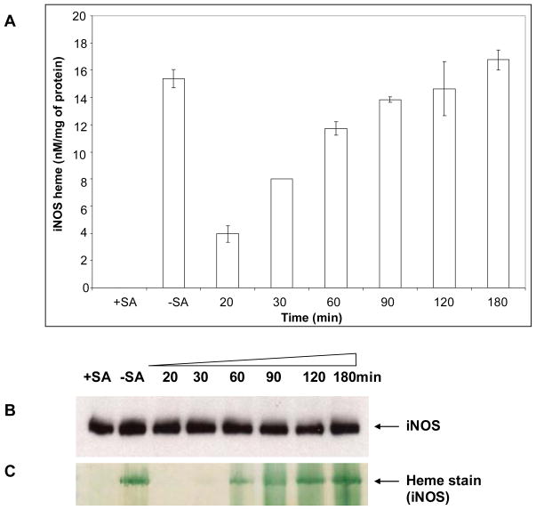
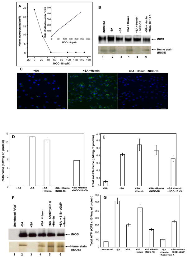
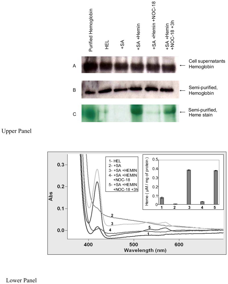
Although the insertion of heme into proteins enables their function in bioenergetics, metabolism, and signaling, the mechanisms and regulation of this process are not fully understood. We developed a means to study cellular heme insertion into apo-protein targets over a 3-h period and then investigated how nitric oxide (NO) released from a chemical donor (NOC-18) might influence heme (protoporphyrin IX) insertion into seven targets that present a range of protein structures, heme ligation states, and functions (three NO synthases, two cytochrome P450's, catalase, and hemoglobin). NO blocked cellular heme insertion into all seven apo-protein targets. The inhibition occurred at relatively low (nM/min) fluxes of NO, was reversible, and did not involve changes in intracellular heme levels, activation of guanylate cyclase, or inhibition of mitochondrial ATP production. These aspects and the range of protein targets suggest that NO can act as a global inhibitor of heme insertion, possibly by inhibiting a common step in the process.


Figures

Figure 1
Figure 1. General method to study heme insertion into heme-free protein targets in cells, and the effect of NO
See text for details.
Figure 2
Figure 2. Cellular heme incorporation into iNOS as a function of added hemin concentration
Raw 264.7 cells were treated with SA for 48 hours and then induced for 16 hours to express apo-iNOS. The cells were then incubated for 3 h with the indicated concentrations of hemin, and soluble cell supernatants were prepared for analysis. Panel A, content of heme-replete iNOS in the cell supernatant samples as determined by difference spectroscopy of the dithionite-reduced, CO-treated samples. Panel B, Western analysis of total iNOS protein expression in the cell supernatant samples that contained equal total protein. Panel C, SDS-PAGE of supernatant samples (equal total protein) followed by in-gel heme staining of the iNOS protein band. Data are representative of 3 separate experiments.
Figure 3
Figure 3. Time course of cellular heme insertion into iNOS
RAW 264.7 cells were cultured and induced to express apo-iNOS as explained in the legend of Fig. 1. Supernatants were made from cell cultures harvested at the indicated times following 7.5 μM hemin addition. Panel A, Buildup of iNOS heme content (determined by the spectroscopic method) versus time, with values normalized per mg total protein. Panel B, Western analysis of total iNOS protein expression in the cell supernatant samples that contained equal total protein. Panel C, In-gel heme staining of the iNOS protein band. Data are representative of 3 separate experiments.
Figure 4
Figure 4. Effect of NO on cellular heme incorporation into iNOS and potential importance of cellular heme uptake, ATP level, or activation of soluble guanylate cyclase
RAW 264.7 cultures with or without 48 h SA pretreatment were induced to express iNOS. Cultures were then treated with 7.5 μM hemin in the absence or presence of the indicated NOC-18 concentrations for 3 h, the cells were harvested and cell supernatants prepared. In some cases the cultures were washed to remove NOC-18 and heme after 3 h and then were cultured an additional 3 h before harvest. In other cases following iNOS induction, RAW 264.7 cells were pretreated for 30 min with Antimycin A (10 μM) or 8-Bromo-cGMP (1mM) prior to hemin addition. Panel A, heme incorporation into iNOS (determined spectroscopically) as a function of NOC-18 concentration. Inset, rates of NO release by NOC-18 under the culture conditions. Panel B, heme incorporation into iNOS under the indicated culture conditions and using 125 μM NOC-18. Panel C, Cellular heme incorporation into iNOS as determined by PIF staining and confocal fluorescence microscopy. SA-pretreated Raw 264.7 cells cultured on glass coverslips were induced to express apo-iNOS and the effect of NOC-18 on heme incorporation was assessed as described above. Confocal images were taken under different conditions as indicated. Panel D, equal total protein contents from each cell supernatant underwent SDS-PAGE analysis and were either heme-stained (the iNOS protein bands are shown) or subject to Western analysis using an anti-iNOS antibody. Panel E, cell supernatants were analyzed for total soluble heme content using the pyridine hemechromogen method. Panel F, equal total protein amounts from each cell supernatant were either Western blotted with iNOS antibody or heme-stained. Panel G, total ATP in the cell supernatants was measured using the ATPlite assay kit, and normalized with respect to the total protein concentrations. Results are representative of two or three trials each.
Figure 5
Figure 5. Effect of NO on cellular heme incorporation in nNOS
HEK293T cell lines stably-expressing nNOS were treated with SA for 48 h followed by 7.5 μM hemin treatment for 3 h in the absence and presence of 125 μM NOC-18. Cells were harvested and cell supernatants prepared. Some cultures treated with hemin and NOC-18 were washed to remove NOC-18 and heme and then cultured for 3 h longer prior to harvesting. Panel A, Heme incorporation into nNOS under the indicated conditions (determined by the spectroscopic method) with values normalized per mg total protein. Panel B, nNOS protein expression in aliquots of equal total protein content for each supernatant sample, and in-gel heme-staining of the nNOS protein bands. Results are representative of three trials.
Figure 6
Figure 6. Effect of NO on cellular heme incorporation in eNOS
Similar experiments were performed with HEK293T cells stably expressing eNOS as described in the legend of Figure 5. Panel A, Heme incorporation into eNOS under the indicated conditions (determined by the spectroscopic method) with values normalized per mg total protein. Panel B, eNOS protein expression in aliquots of equal total protein content for each supernatant sample, and in-gel heme-staining of the eNOS protein bands. Results are representative of three trials.
Figure 7
Figure 7. Effect of NO on cellular heme incorporation into CYP 2D6 and 3A4
HEK293T cells were treated with SA for 48 h followed by transient transfection to initiate CYP 2D6 or 3A4 protein expression. At 24 h post-transfection the cells were treated with 7.5 μM hemin alone or with 125 μM NOC-18 for 3 h before being harvested and supernatants prepared. Some cell cultures were washed to remove NOC-18 and hemin and were then cultured for an additional 3 h prior to harvesting. Panel A, Heme incorporation into the CYP apo-enzymes under the indicated conditions (determined by the spectroscopic method) with values normalized per mg total protein. Panels B and C, Western analysis of CYP protein expression levels in aliquots of equal total protein content for the supernatant samples, and corresponding in-gel heme staining of the CYP protein bands. Results are representative of three trials.
Figure 8
Figure 8. Effect of NO on cellular heme incorporation into catalase
HEK293T cells were treated with SA for 48 h followed by transient transfection of vector for catalase expression. At 24 h post-transfection the cells were treated with 7.5 μM hemin alone or plus 125 μM NOC-18 and were incubated for 3 h before being harvested. Some of the cultures had their media replaced to study hemin incorporation for 3 h after NOC-18 removal. (A) Western analysis of catalase expression in aliquots of 250μg total protein from each indicated supernatant sample, and corresponding in-gel heme staining of the catalase bands. (B) Catalase activity determined for aliquots of equal total protein from the indicated supernatant samples. Results are representative of three trials.
Figure 9
Figure 9. Effect of NO on cellular heme insertion into hemoglobin
Human erythroid leukemia (HEL) cell line K562 were treated with SA for 72 h and then the cells were given 7.5 μM hemin alone or plus 125 μM NOC-18 and were incubated for 3 h before being harvested. Some of the cultures had their media replaced to study hemin incorporation for 3 h after NOC-18 removal. Upper panels- (A) Western analyses of hemoglobin expression in equal total protein amounts of each cell supernatant as indicated or (B) hemoglobin levels in the semi-purified supernatant samples, (C) the corresponding in-gel heme stain of the semi-purified hemoglobin protein bands. Lower panel- UV-visible spectra of semi-purified hemoglobin fractions from the various supernatants as indicated, depicting the Soret absorption peak at 414 nm. Inset shows a bar graph depicting the heme content of the semi-purified hemoglobin fractions based on the Soret abrobance at 414 nm. Results are representative of three trials.

Similar articles

Cited by

References

    1. Raven E, Ortiz de Montellano PR. Editorial: The chemistry and biochemistry of heme proteins. Nat Prod Rep. 2007;24:499.
    1. Paoli M, Marles-Wright J, Smith A. Structure-function relationships in heme-proteins. DNA Cell Biol. 2002;21:271–280. - PubMed
    1. Milo R, Hou JH, Springer M, Brenner MP, Kirschner MW. The relationship between evolutionary and physiological variation in hemoglobin. Proc Natl Acad Sci U S A. 2007;104:16998–7003. - PMC - PubMed
    1. Poulos TL. Structural biology of heme monooxygenases. Biochem Biophys Res Commun. 2005;338:337–345. - PubMed
    1. Zhu Y, Silverman RB. Revisiting heme mechanisms. A perspective on the mechanisms of nitric oxide synthase (NOS), Heme oxygenase (HO), and cytochrome P450s (CYP450s) Biochemistry. 2008;47:2231–2243. - PubMed

Publication types

LinkOut - more resources