

Human intestinal MUC17 mucin augments intestinal cell restitution and enhances healing of experimental colitis
- PMID: 20211273
- PMCID: PMC2919344
- DOI: 10.1016/j.biocel.2010.03.001
Human intestinal MUC17 mucin augments intestinal cell restitution and enhances healing of experimental colitis
Abstract
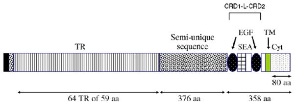
The membrane-bound mucins, MUC17 (human) and Muc3 (mouse), are highly expressed on the apical surface of intestinal epithelia and are thought to be cytoprotective. The extracellular regions of these mucins contain EGF-like Cys-rich segments (CRD1 and CRD2) connected by an intervening linker domain (L). The purpose of this study was to determine the functional activity of human MUC17 membrane-bound mucin.
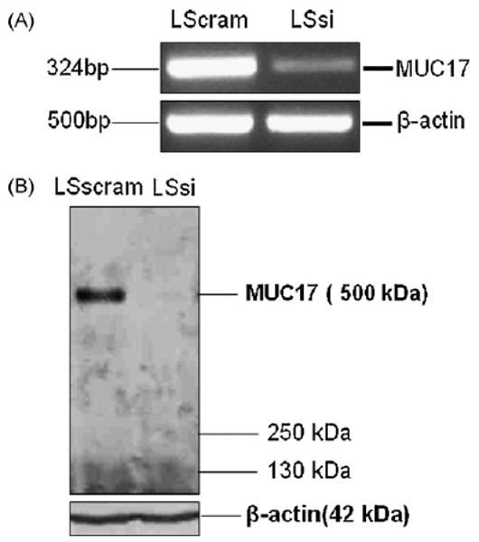
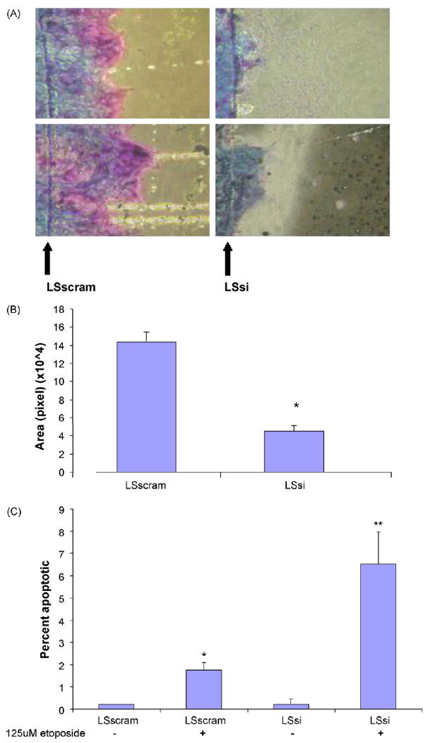
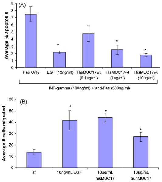
Methods: Endogenous MUC17 was inhibited in LS174T colon cells by stable transfection of a small hairpin RNA targeting MUC17 (LSsi cells). The effect of recombinant MUC17-CRD1-L-CRD2 protein on migration, apoptosis, and experimental colitis was determined.
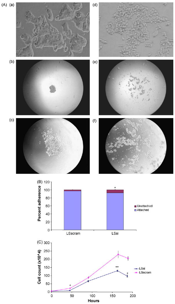
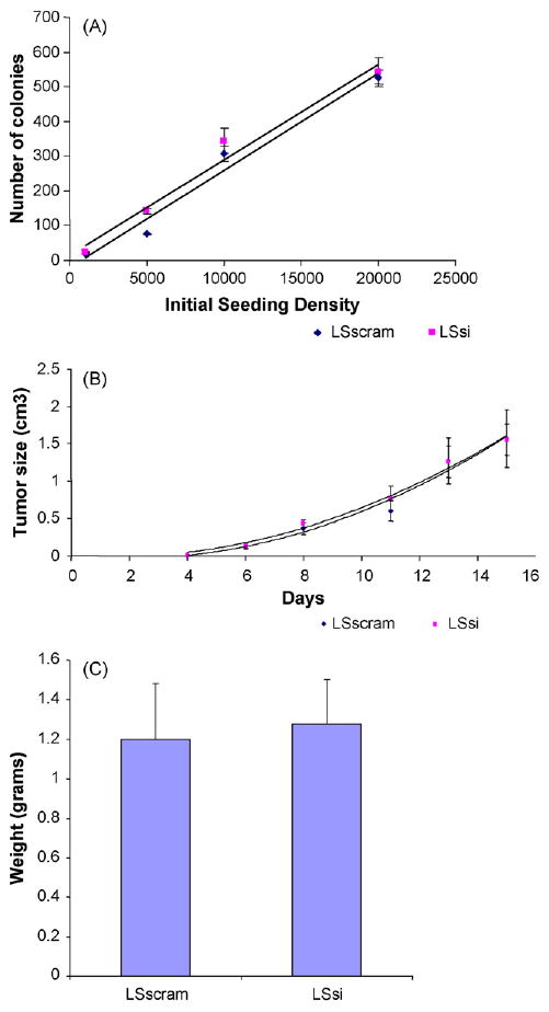
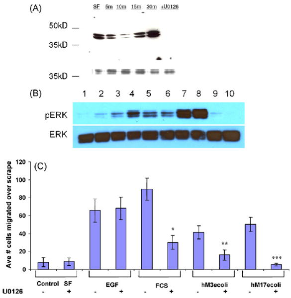
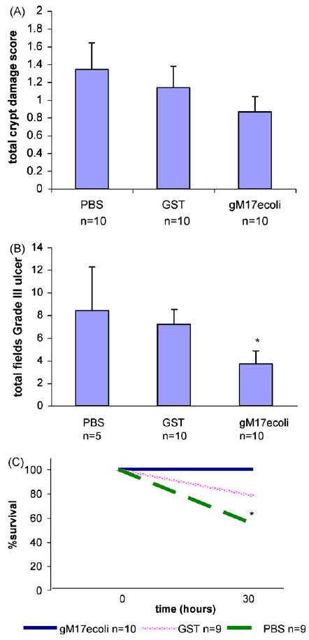
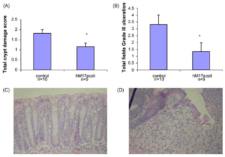
Results: Reduced MUC17 expression in LSsi cells was associated with visibly reduced cell aggregation, reduced cell-cell adherence, and reduced cell migration, but no change in tumorigenicity. LSsi cells also demonstrated a 3.7-fold increase in apoptosis rates compared with control cells following treatment with etoposide. Exposure of colonic cell lines to exogenous recombinant MUC17-CRD1-L-CRD2 protein significantly increased cell migration and inhibited apoptosis. As a marker of biologic activity, MUC17-CRD1-L-CRD2 proteins stimulate ERK phosphorylation in colonic cell lines; and inhibition of ERK phosphorylation reduced the anti-apoptosis and migratory effect of MUC17-CRD1-L-CRD2. Finally, mice treated with MUC17-CRD1-L-CRD2 protein given per rectum demonstrated accelerated healing in acetic acid and dextran sodium sulfate induced colitis in vivo. These data indicate that both native MUC17 and the exogenous recombinant cysteine-rich domain of MUC17 play a role in diverse cellular mechanisms related to cell restitution, and suggest a potential role for MUC17-CRD1-L-CRD2 recombinant protein in the treatment of mucosal inflammatory diseases.
Published by Elsevier Ltd.
Figures
Similar articles
-
Expression of intestinal MUC17 membrane-bound mucin in inflammatory and neoplastic diseases of the colon.J Clin Pathol. 2010 Aug;63(8):702-7. doi: 10.1136/jcp.2010.078717. J Clin Pathol. 2010. PMID: 20702471 Free PMC article.
-
Activity of recombinant cysteine-rich domain proteins derived from the membrane-bound MUC17/Muc3 family mucins.Biochim Biophys Acta. 2010 Jul;1800(7):629-38. doi: 10.1016/j.bbagen.2010.03.010. Epub 2010 Mar 20. Biochim Biophys Acta. 2010. PMID: 20332014 Free PMC article.
-
Muc17 protects intestinal epithelial cells from enteroinvasive E. coli infection by promoting epithelial barrier integrity.Am J Physiol Gastrointest Liver Physiol. 2011 Jun;300(6):G1144-55. doi: 10.1152/ajpgi.00138.2010. Epub 2011 Mar 10. Am J Physiol Gastrointest Liver Physiol. 2011. PMID: 21393431 Free PMC article.
-
Cysteine-rich domains of muc3 intestinal mucin promote cell migration, inhibit apoptosis, and accelerate wound healing.Gastroenterology. 2006 Nov;131(5):1501-17. doi: 10.1053/j.gastro.2006.09.006. Epub 2006 Sep 9. Gastroenterology. 2006. PMID: 17101324
-
Intestinal goblet cells and mucins in health and disease: recent insights and progress.Curr Gastroenterol Rep. 2010 Oct;12(5):319-30. doi: 10.1007/s11894-010-0131-2. Curr Gastroenterol Rep. 2010. PMID: 20703838 Free PMC article. Review.
Cited by
-
Enhanced membrane-tethered mucin 3 (MUC3) expression by a tetrameric branched peptide with a conserved TFLK motif inhibits bacteria adherence.J Biol Chem. 2013 Feb 22;288(8):5407-16. doi: 10.1074/jbc.M112.408245. Epub 2013 Jan 10. J Biol Chem. 2013. PMID: 23316049 Free PMC article.
-
MUC17 Is a Potential New Prognostic Biomarker and Promotes Pancreatic Cancer Progression in Obstructive Jaundice.Oncology. 2025;103(8):725-741. doi: 10.1159/000541874. Epub 2024 Oct 11. Oncology. 2025. PMID: 39396510 Free PMC article.
-
Expression of intestinal MUC17 membrane-bound mucin in inflammatory and neoplastic diseases of the colon.J Clin Pathol. 2010 Aug;63(8):702-7. doi: 10.1136/jcp.2010.078717. J Clin Pathol. 2010. PMID: 20702471 Free PMC article.
-
Epithelial restitution defect in neonatal jejunum is rescued by juvenile mucosal homogenate in a pig model of intestinal ischemic injury and repair.PLoS One. 2018 Aug 23;13(8):e0200674. doi: 10.1371/journal.pone.0200674. eCollection 2018. PLoS One. 2018. PMID: 30138372 Free PMC article.
-
Membrane-bound mucins and mucin terminal glycans expression in idiopathic or Helicobacter pylori, NSAID associated peptic ulcers.Dig Dis Sci. 2012 Oct;57(10):2535-44. doi: 10.1007/s10620-012-2205-5. Epub 2012 May 11. Dig Dis Sci. 2012. Retraction in: Dig Dis Sci. 2013 Jun;58(6):1812. doi: 10.1007/s10620-013-2723-9. PMID: 22576713 Retracted.
References
-
- Argenzio RA. Comparative pathophysiology of nonglandular ulcer disease: a review of experimental studies. Equine Vet J Suppl. 1999;29:19–23. - PubMed
-
- Chaturvedi P, Singh AP, Moniaux N, Senapati S, Chakraborty S, Meza JL, et al. MUC4 mucin potentiates pancreatic tumor cell proliferation, survival, and invasive properties and interferes with its interaction to extracellular matrix proteins. Mol Cancer Res. 2007;5(4):309–20. - PubMed
-
- Cooper HS, Murthy SN, Shah RS, Sedergran DJ. Clinicopathologic study of dextran sulfate sodium experimental murine colitis. Lab Invest. 1993;69(2):238–49. - PubMed
Publication types
MeSH terms
Substances
Grants and funding
LinkOut - more resources
Full Text Sources
Other Literature Sources
Research Materials
Miscellaneous
