. 2010 Jun;38(11):3834-47.
doi: 10.1093/nar/gkq124. Epub 2010 Mar 7.

Direct observation of the temperature-induced melting process of the Salmonella fourU RNA thermometer at base-pair resolution

Affiliations

Direct observation of the temperature-induced melting process of the Salmonella fourU RNA thermometer at base-pair resolution

Jörg Rinnenthal et al. Nucleic Acids Res. 2010 Jun.

Abstract

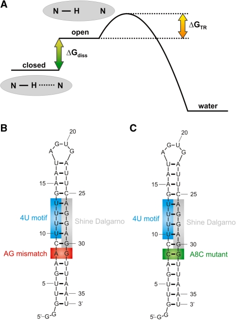
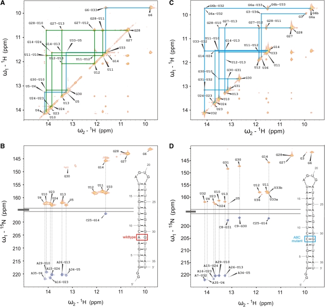
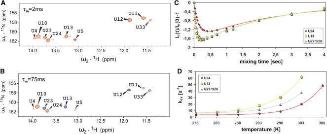
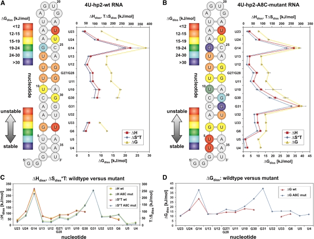
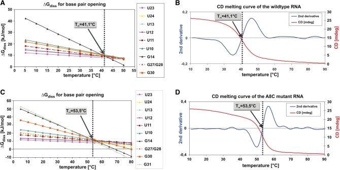
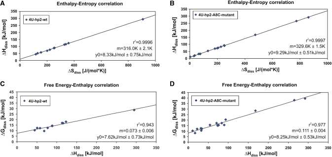
In prokaryotes, RNA thermometers regulate a number of heat shock and virulence genes. These temperature sensitive RNA elements are usually located in the 5'-untranslated regions of the regulated genes. They repress translation initiation by base pairing to the Shine-Dalgarno sequence at low temperatures. We investigated the thermodynamic stability of the temperature labile hairpin 2 of the Salmonella fourU RNA thermometer over a broad temperature range and determined free energy, enthalpy and entropy values for the base-pair opening of individual nucleobases by measuring the temperature dependence of the imino proton exchange rates via NMR spectroscopy. Exchange rates were analyzed for the wild-type (wt) RNA and the A8C mutant. The wt RNA was found to be stabilized by the extraordinarily stable G14-C25 base pair. The mismatch base pair in the wt RNA thermometer (A8-G31) is responsible for the smaller cooperativity of the unfolding transition in the wt RNA. Enthalpy and entropy values for the base-pair opening events exhibit linear correlation for both RNAs. The slopes of these correlations coincide with the melting points of the RNAs determined by CD spectroscopy. RNA unfolding occurs at a temperature where all nucleobases have equal thermodynamic stabilities. Our results are in agreement with a consecutive zipper-type unfolding mechanism in which the stacking interaction is responsible for the observed cooperativity. Furthermore, remote effects of the A8C mutation affecting the stability of nucleobase G14 could be identified. According to our analysis we deduce that this effect is most probably transduced via the hydration shell of the RNA.


Figures

Figure 1.
Figure 1.
(A) Energy diagram describing the imino proton exchange process from the nucleobase in the closed (base paired) conformation to the water. (B) Secondary structure of the 4U-hp2-wt RNA (C) Secondary structure of the 4U-hp2-A8C-mutant RNA. The A8C mutant corresponds to the A29C mutant of the full-length RNA (17).
Figure 2.
Figure 2.
(A) Imino region of a 1H-1H-NOESY spectrum recorded on a 4U-hp2-wt RNA sample with a mixing time Tm of 150 ms. Two separate sequential walks are indicated with blue (U4–G6) and green (G30–U23) lines. Assignment labels of cross and diagonal peaks are shown in black. (B) HNN-COSY spectrum recorded on the 4U-hp2-wt RNA. Additionally, the secondary structure of the 4U-hp2-wt RNA is illustrated within the spectrum. Diagonal peaks are given in orange, cross peaks in blue. The corresponding cross and diagonal peaks are connected by dotted lines. (C) Imino region of a 1H-1H-NOESY spectrum recorded on a 4U-hp2-A8C-mutant RNA sample with a mixing time Tm of 150 ms. Sequential walk is indicated in blue. Assignment labels of cross and diagonal peaks are shown in black. (D) HNN-COSY spectrum recorded on the 4U-hp2-A8C-mutant RNA. Additionally, the secondary structure of the 4U-hp2-A8C mutant RNA is illustrated within the spectrum. Diagonal peaks are given in orange, cross peaks in blue. The corresponding cross and diagonal peaks are connected by dotted lines.
Figure 3.
Figure 3.
1H-15N-2D-slice of the Uracil imino resonances of the 15N-edited inversion recovery experiment recorded on a 4U-hp2-wt RNA sample (A) at a temperature of 20°C and a mixing time of τm = 2 ms. Positive signals are illustrated in orange and (B) at a temperature of 20°C and a mixing time of τm = 75 ms. Positive signals are illustrated in orange, negative signals in blue. (C) Dependence of the intensities of the imino signals U24 (red diamonds), U13 (yellow squares) and G27/G28 (blue triangles) on the mixing time τm. The corresponding fits according to Equation (2) are indicated as solid lines. (D) Temperature dependence of the imino proton exchange rates kex of U24 (red diamonds), U13 (yellow squares) and G27/G28 (blue triangles). The corresponding fits according to Equation (14) are indicated as solid lines.
Figure 4.
Figure 4.
(A) Left: free energy representation for the base-pair opening events for the imino resonances belonging to a particular nucleobase of the 4U-hp2-wt-RNA. Red color corresponds to low stability of the closed state with respect to the open state while blue indicates high stability of the closed state in comparison to the open state. Right: diagram representation showing the ΔGdiss (20°C), ΔHdiss, and ΔSdiss*T (20°C) values for the base-pair opening of individual nucleobases of the 4U-hp2-wt RNA (B) Same representation as in (A) for the 4U-hp2-A8C-mutant RNA. (C) Diagram comparing the ΔHdiss and ΔSdiss*T (20°C) values of the 4U-hp2-wt RNA and the 4U-hp2-A8C-mutant RNA. ΔHdiss and ΔSdiss*T (20°C) values are plotted on different scales. 4U-hp2-wt: ΔHdiss (yellow squares), ΔSdiss*T at T = 20°C (red triangles). 4U-hp2-A8C-mutant: ΔHdiss (green circles), ΔSdiss*T at T = 20°C (blue diamonds). (D) Diagram comparing the ΔGdiss (20°C) values of the 4U-hp2-wt RNA (red triangles) and the 4U-hp2-A8C-mutant RNA (blue diamonds).
Figure 5.
Figure 5.
(A) ΔGdiss(T) for the base-pair opening event of individual nucleobases in the 4U-hp2-wt RNA. ΔGdiss(T) values are derived from the corresponding ΔHdiss and ΔSdiss values according to the Gibbs–Helmholtz equation (Equation 11) and are extrapolated to higher temperatures. Additionally, the melting point Tm of the 4U-hp2-wt-RNA according to the CD melting curve is indicated. (B) The CD melting curve of the 4U-hp2-wt RNA recorded at a wavelength of 258 nm is depicted as red line. Second derivative of the CD melting curve is shown as blue line. (C) Same representation as (A) but for the 4U-hp2-A8C-mutant RNA (D) Same representation as (B) but for the 4U-hp2-A8C-mutant RNA.
Figure 6.
Figure 6.
(A) Enthalpy–entropy correlation for the base-pair opening event of individual nucleobases in the 4U-hp2-wt RNA. (B) Enthalpy–entropy correlation for the base-pair opening event of individual nucleobases in the 4U-hp2-A8C-mutant RNA. (C) Free energy–entropy correlation for the base-pair opening event of individual nucleobases in the 4U-hp2-wt RNA. (D) Free energy–entropy correlation for the base-pair opening event of individual nucleobases in the 4U-hp2-A8C-mutant RNA. Plots were fitted according to the linear equation f = y0 + mx. Linear fitting results are illustrated within the figures.

Similar articles

Cited by

References

    1. Bocobza SE, Aharoni A. Switching the light on plant riboswitches. Trends Plant Sci. 2008;13:526–533. - PubMed
    1. Sudarsan N, Barrick JE, Breaker RR. Metabolite-binding RNA domains are present in the genes of eukaryotes. RNA. 2003;9:644–647. - PMC - PubMed
    1. Croft MT, Moulin M, Webb ME, Smith AG. Thiamine biosynthesis in algae is regulated by riboswitches. Proc. Natl Acad. Sci. USA. 2007;104:20770–20775. - PMC - PubMed
    1. Sharp PA. The centrality of RNA. Cell. 2009;136:577–580. - PubMed
    1. Waters LS, Storz G. Regulatory RNAs in bacteria. Cell. 2009;136:615–628. - PMC - PubMed

Publication types

Substances