Review
. 2010 Jul;51(7):1643-75.
doi: 10.1194/jlr.R003996. Epub 2010 Mar 8.

Multi-system disorders of glycosphingolipid and ganglioside metabolism

Affiliations
Review

Multi-system disorders of glycosphingolipid and ganglioside metabolism

You-Hai Xu et al. J Lipid Res. 2010 Jul.

Abstract

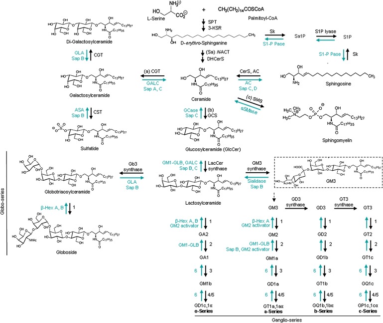
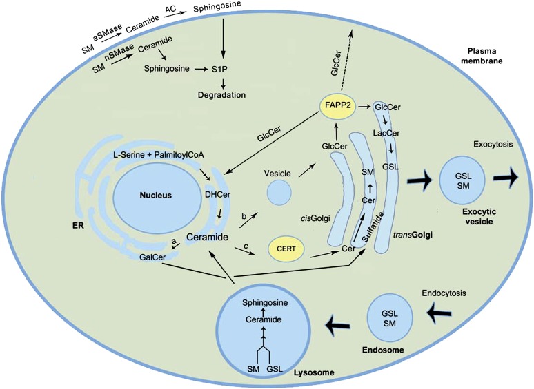
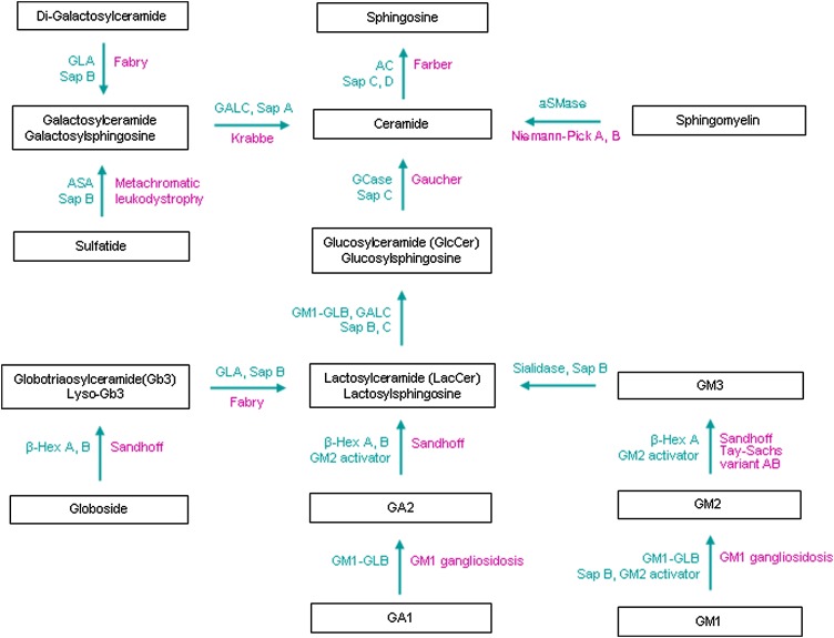
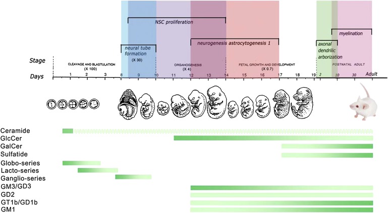
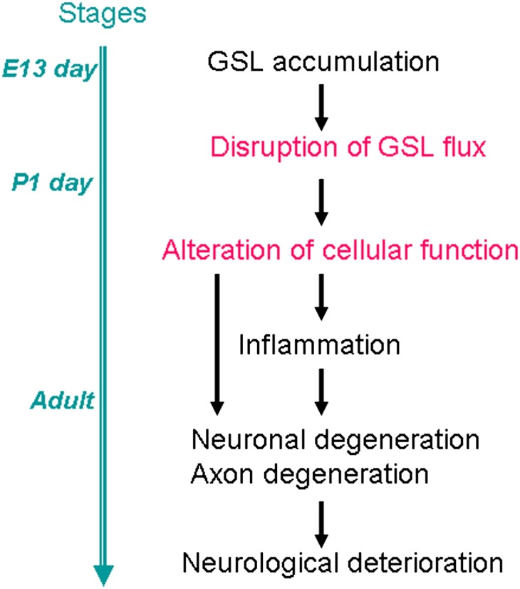
Glycosphingolipids (GSLs) and gangliosides are a group of bioactive glycolipids that include cerebrosides, globosides, and gangliosides. These lipids play major roles in signal transduction, cell adhesion, modulating growth factor/hormone receptor, antigen recognition, and protein trafficking. Specific genetic defects in lysosomal hydrolases disrupt normal GSL and ganglioside metabolism leading to their excess accumulation in cellular compartments, particularly in the lysosome, i.e., lysosomal storage diseases (LSDs). The storage diseases of GSLs and gangliosides affect all organ systems, but the central nervous system (CNS) is primarily involved in many. Current treatments can attenuate the visceral disease, but the management of CNS involvement remains an unmet medical need. Early interventions that alter the CNS disease have shown promise in delaying neurologic involvement in several CNS LSDs. Consequently, effective treatment for such devastating inherited diseases requires an understanding of the early developmental and pathological mechanisms of GSL and ganglioside flux (synthesis and degradation) that underlie the CNS diseases. These are the focus of this review.


Figures

Fig. 1.
Fig. 1.
Schematic view of the GSL metabolism pathways. The synthesis of GSLs and gangliosides progress stepwise and are catalyzed by membranous glycosyltransferases in the ER or Golgi apparatus (see text). The degradation reactions are also sequential and occur within the lysosomes by various hydrolases. The black arrows show the synthesis pathway in the ER and Golgi and the green arrows show the degradation pathway in the lysosome. The enzymes in o-, a-, b-, and c-ganglioside series are numbered: 1. GM2/GD2/GA2/GT2-synthase (β-1,4-N-acetyl-galactosaminyltransferase, GalNacT), 2. GA1/GM1a/GD1b/GT1c-synthase (UDP-Gal:βGalNAc β-1,3-galactosyltransferase), 3. sialyltransferase IV, 4. sialyltransferase V, 5. sialyltransferase VII, 6. sialidase. Abbreviations: 3-KSR (3-ketosphinganine reductase), Sk (sphingosine kinase), Sa1P (sphinganine 1-phosphate), S1P (sphingosine 1-phosphate), S1-P Pase (sphingosine 1-phosphate phosphatase), (Sa) N-ACT (sphinganine N-acyltransferase), DHCerS (dihydroceramide desaturase), CerS (ceramide synthase, also called longevity assurance genes), SMS (sphingomyelin synthase), GT3 synthase (α-N-acetyl-neuraminide α-2,8- sialyltransferase). The chemical structures are adapted from (, , , –315) and http://www.cybercolloids.net/library/sugars/hexoses.php. Nomenclature of the enzymes and protein are from IUBMB (http://www.chem.qmul.ac.uk/iubmb/enzyme) (37, 43, 315).
Fig. 2.
Fig. 2.
Intracellular topology of GSL biosynthesis and trafficking. Ceramide, formed by condensation of serine and palmitoyl-CoA on the cytoplasmic face of ER, has one of three fates (16): a) conversion to galactosylceramide (GalCer) in the ER lumen, which is subsequently converted to sulfatide in the mid-Golgi (13, 14), b) vesicular transport to the cytoplasmic face of the cis-Golgi where it is a precursor for GlcCer synthesis (26, 316), and c) transport by ceramide transfer protein (CERT) to the mid-Golgi where sphingomyelin is formed within the lumen (319). Once formed, GlcCer also has several fates (27, 317): 1) transport by FAPP2 to the ER and/or to the trans-Golgi lumen where it is converted to lactosylceramide (LacCer) (17, 26, 27), 2) to the cytoplasmic side of the plasma membrane by unknown mechanisms, and 3) to the extracellular matrix by exocytosis. Addition of sialic acid to LacCer initiates synthesis of gangliosides or the sialo-GSL series (318). Ceramide can also be generated through degradation of sphingomyelin in the lysosome or at the plasma membrane by aSMase and nSMase (10). Newly synthesized GSLs can exit the cell by exocytic vesicles while membrane and extracellular GSLs can be transported intracellularly via endocytosis with subsequent degradation sphingosine and free fatty acids by hydrolases in the lysosome (2, 10). Abbreviations: DHCer (dihydroceramide), S1P (sphingosine 1-phosphate), SM (sphingomyelin).
Fig. 3.
Fig. 3.
Disorders of GSL and ganglioside degradation. Inherited diseases (violet) caused by genetic defects of individual hydrolases/proteins (green) in the GSL and ganglioside degradation pathway. Increased levels of lysosphingolipids occur in the GSL LSDs, e.g., glucosylsphingosine in Gaucher disease, Lyso-Gb3 in Fabry disease, and galactosylsphingosine in Krabbe disease. Variant AB is GM2 activator deficiency disease. Tay-Sach disease, Sandhoff disease, and Variant AB are GM2 gangliosidosis. Abbreviations are listed in Fig. 1 legend.
Fig. 4.
Fig. 4.
GSLs and gangliosides in developmental stages of the nervous system. The diagram shows GSL profiles during development of the mouse nervous system. Mouse embryonic developmental stages are from single cell (∼ day 1), 16 cells (∼ day 3) to fetal (day 19). The mouse embryonic and neural developmental stages are described (320). Colored (vertical) bars above the timeline illustrate overlapping developmental stages. The horizontal gradient bars show the dynamic changes of GSL and ganglioside levels that occur during the stages of mouse embryo and neural development based upon experimental observations (22, 39, 40, 43, 44) (Sun et al., unpublished observations). The dark green indicates increased levels of the specific lipids and the less dense green implies lower levels. Ceramide could be identified at 2-4 cell stage (39). The hatched bar demonstrates the hypothetical ceramide levels since ceramide synthesis remains active throughout life.
Fig. 5.
Fig. 5.
Hypothetical scheme for neuronal disease progression in the nervous system. Accumulation of GSLs causes abnormalities of neuronal cell functions in mouse models and the human diseases. Abnormal degradation of GSL may disrupt the normal flux of GSL in the cell, which can affect a variety of cellular functions. How the GSL accumulation alters GSL flux and cellular function is largely unknown. The changes of the cellular function may cause neuronal degeneration. Inflammation is often detected before the neuronal and axonal defects and it plays a role in potentiating disease progression. The blue line on the left shows the stage of brain development.

Similar articles

Cited by

References

    1. Holthuis J. C., Pomorski T., Raggers R. J., Sprong H., Van Meer G. 2001. The organizing potential of sphingolipids in intracellular membrane transport. Physiol. Rev. 81: 1689–1723. - PubMed
    1. Kolter T., Sandhoff K. 2005. Principles of lysosomal membrane digestion: stimulation of sphingolipid degradation by sphingolipid activator proteins and anionic lysosomal lipids. Annu. Rev. Cell Dev. Biol. 21: 81–103. - PubMed
    1. Lahiri S., Futerman A. H. 2007. The metabolism and function of sphingolipids and glycosphingolipids. Cell. Mol. Life Sci. 64: 2270–2284. - PMC - PubMed
    1. Mandon E. C., Ehses I., Rother J., van Echten G., Sandhoff K. 1992. Subcellular localization and membrane topology of serine palmitoyltransferase, 3-dehydrosphinganine reductase, and sphinganine N-acyltransferase in mouse liver. J. Biol. Chem. 267: 11144–11148. - PubMed
    1. Michel C., van Echten-Deckert G., Rother J., Sandhoff K., Wang E., Merrill A. H., Jr 1997. Characterization of ceramide synthesis. A dihydroceramide desaturase introduces the 4,5-trans-double bond of sphingosine at the level of dihydroceramide. J. Biol. Chem. 272: 22432–22437. - PubMed

Publication types

MeSH terms

LinkOut - more resources