. 2010 Jun;115(2):422-34.
doi: 10.1093/toxsci/kfq063. Epub 2010 Mar 8.

Effects of TCDD on the fate of naive dendritic cells

Affiliations

Effects of TCDD on the fate of naive dendritic cells

Jaishree Bankoti et al. Toxicol Sci. 2010 Jun.

Abstract

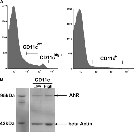
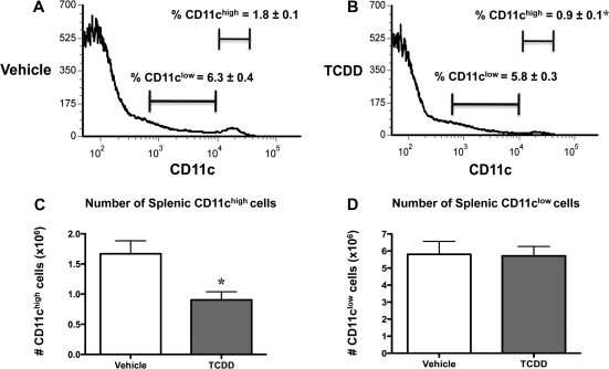
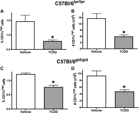
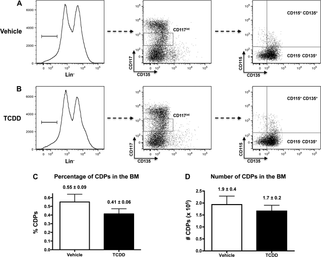
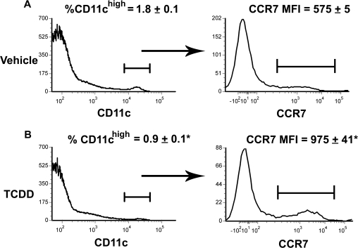
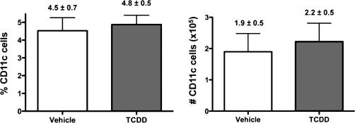
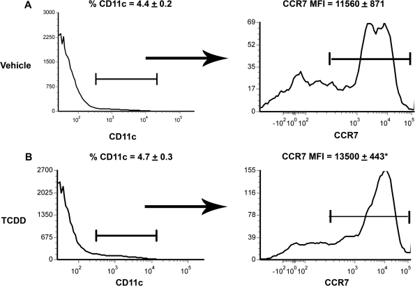
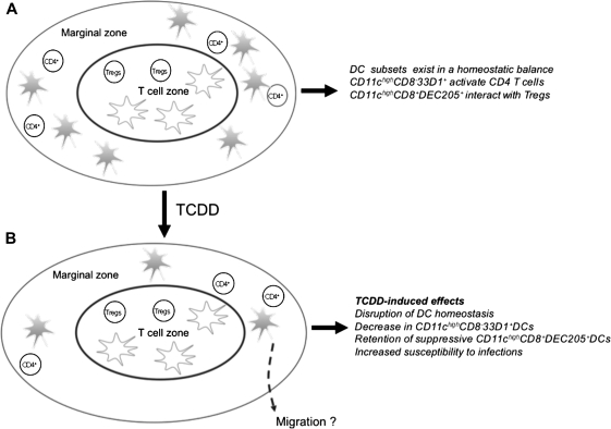
The environmental contaminant, 2,3,7,8-tetrachlorodibenzo-p-dioxin (TCDD), causes immune suppression via activation of the aryl hydrocarbon receptor. Dendritic cells (DCs), the professional antigen-presenting cells in the immune system, are adversely affected by TCDD. We hypothesized that TCDD alters DC homeostasis, resulting in a loss of DCs in naive mice. To test this hypothesis, C57Bl/6 mice were gavaged with either vehicle or an immunosuppressive dose of TCDD (15 microg/kg). TCDD exposure decreased the frequency and number of splenic CD11c(high) DCs on day 7 when compared with vehicle-treated controls. TCDD increased the expression of CD86 and CD54, while decreasing the frequency of splenic CD11c(high) DCs expressing CD11a and major histocompatibility complex (MHC) class II. Moreover, TCDD selectively decreased the CD11c(high)CD8alpha(-)33D1(+) splenic DCs specialized at activating CD4(+) T cells but did not affect the regulatory CD11c(high)CD8alpha(+)DEC205(+) splenic DCs. TCDD did not alter the number or frequency of CD11c(low) splenic DCs but decreased their MHC class II and CD11a expression. Loss of splenic CD11c(high) DCs was independent of Fas-mediated apoptosis and was not due to alterations in the numbers of common DC precursors in the bone marrow or their ability to generate steady-state DCs in vitro. Instead, increased CCR7 expression on CD11c(high) DCs suggested involvement of a migratory event. Popliteal and brachial lymph node CD11c(+) cells showed elevated levels of MHC class II and CD40 following TCDD exposure. Collectively, this study shows the presence of a TCDD-sensitive splenic DC subpopulation in naive mice, suggesting that TCDD may induce suppression of T-cell-mediated immunity by disrupting DC homeostasis.


Figures

FIG. 1.
FIG. 1.
(A) CD11c expression on DCs from peripheral immune tissues. Histograms represent two distinct populations of CD11chigh and CD11c low DCs in the splenic tissue of unimmunized mice and a homogenous population of CD11c+ DCs in the LNs. (B) AhR protein levels in splenic CD11chigh and CD11clow DCs. Lysates were prepared from a single Flt3 ligand–treated C57Bl/6 mouse and a single experiment. Spleens were harvested on day 11, and DC lysates were prepared from FACS-sorted CD11chigh and CD11clow DCs. Purified nonadherent BMDCs were used as a positive control for the expression of AhR. Whole cell lysates were evaluated by Western blotting for AhR and β-actin as described in the “Materials and Methods” section.
FIG. 2.
FIG. 2.
TCDD induces a loss of splenic CD11chigh but not CD11clow DCs. C57Bl/6 mice were treated with vehicle or TCDD (15 μg/kg). Spleens were harvested on day 7, processed, and phenotyped by flow cytometry as described in the “Materials and Methods” section. Histograms represent the percentage of splenic CD11chigh and CD11clow DCs from vehicle- (A) and TCDD-treated (B) mice. Bar graphs represent the total number of splenic CD11chigh (C) and CD11clow (D) DCs from vehicle- and TCDD-treated mice. Data represent mean ± SEM of six mice per treatment group and are representative of four independent experiments. *p ≤ 0.05 for the comparison between vehicle- and TCDD-treated mice.
FIG. 3.
FIG. 3.
The number and percentage of splenic CD11chigh DCs in Fas- and FasL-deficient mice are decreased following TCDD exposure. Nonimmunized C57Bl/6lpr/lpr (A and B) and C57Bl/6gld/gld mice (C and D) were treated with vehicle or TCDD (15 μg/kg) and the numbers of resident DCs in the spleen were determined on day 7. Data represent the mean ± SEM of five to six mice per treatment group and are representative of two independent experiments for C57Bl/6lpr/lpr and one experiment for C57Bl/6gld/gld mice. *p ≤ 0.05 for the comparison between vehicle- and TCDD-treated mice.
FIG. 4.
FIG. 4.
TCDD does not affect CDP levels in the BM. On day 7 following exposure, BM cells were harvested from vehicle- or TCDD-treated C57Bl/6 mice and CDPs analyzed by flow cytometry as described in the “Materials and Methods” section. Representative histograms show the LinCD117intCD135+CD115+ CDPs from vehicle- (A) and TCDD-treated (B) mice and the lack of effects of TCDD on the frequencies (C) and numbers (D) of CDPs in the BM. Values shown are the mean ± SEM of five to six mice per treatment and are representative of two independent experiments.
FIG. 5.
FIG. 5.
TCDD increases CCR7 expression on splenic CD11chigh DCs. C57Bl/6 mice were treated with vehicle or TCDD (15 μg/kg), and spleens were harvested and processed for flow cytometric analysis on day 5 as described in the “Materials and Methods” section. Representative histograms represent mean fluorescence intensity (MFI) of CCR7 expression on CD11chigh cells from vehicle- (A) and TCDD-treated (B) mice. Values shown are mean ± SEM for four to five mice per treatment group and are representative of two independent experiments. *p ≤ 0.05 for the comparison between vehicle- and TCDD-treated mice.
FIG. 6.
FIG. 6.
Effects of TCDD on DCs in the peripheral LNs. The percentages and numbers of CD11c+ DCs in the PBLNs were determined from mice treated with TCDD (15 μg/kg) or vehicle for 7 days. LNs were harvested and DCs analyzed by flow cytometry as described in the “Materials and Methods” section. Data represent the mean ± SEM of four to six mice per treatment group and are representative of three independent experiments.
FIG. 7.
FIG. 7.
Expression of CCR7 is increased on DCs from LNs following TCDD exposure. On day 5 following exposure to vehicle or TCDD, LNs were harvested and analyzed by flow cytometric analysis as previously described. Representative histograms depict the mean fluorescence intensity (MFI) of CCR7 expression on CD11c+ cells in mice treated with (A) vehicle and (B) TCDD. Values represent the mean ± SEM of five mice per treatment group and are representative of two independent experiments. *p ≤ 0.05 for the comparison between vehicle- and TCDD-treated mice.
FIG. 8.
FIG. 8.
Schematic diagram representing the effects of TCDD on splenic CD11chigh DCs. (A) In naive mice, splenic CD11chigh DC subsets exist in a homeostatic balance. The CD11chighCD8α33D1+ DCs (shown in gray) reside in the marginal zone and red pulp and generate effector CD4+ T cells. The CD11chighCD8α+DEC205+ DCs reside in the T-cell zone and are specialized at inducing/maintaining Tregs. (B) Following TCDD exposure, DC homeostasis is perturbed in the spleen. TCDD induces a loss of CD11chighCD8α33D1+ DCs and retention of Treg-inducing CD11chighCD8α+DEC205+ DCs.

Similar articles

Cited by

References

    1. Adachi M, Watanabe-Fukunaga R, Nagata S. Aberrant transcription caused by the insertion of an early transposable element in an intron of the Fas antigen gene of lpr mice. Proc. Natl. Acad. Sci. U.S.A. 1993;90:1756–1760. - PMC - PubMed
    1. Banchereau J, Schuler-Thurner B, Palucka AK, Schuler G. Dendritic cells as vectors for therapy. Cell. 2001;106:271–274. - PubMed
    1. Brasel K, De Smedt T, Smith JL, Maliszewski CR. Generation of murine dendritic cells from flt3-ligand-supplemented bone marrow cultures. Blood. 2000;96:3029–3039. - PubMed
    1. Brawand P, Fitzpatrick DR, Greenfield BW, Brasel K, Maliszewski CR, De Smedt T. Murine plasmacytoid pre-dendritic cells generated from Flt3 ligand-supplemented bone marrow cultures are immature APCs. J. Immunol. 2002;169:6711–6719. - PubMed
    1. Camacho IA, Hassuneh MR, Nagarkatti M, Nagarkatti PS. Enhanced activation-induced cell death as a mechanism of 2,3,7,8-tetrachlorodibenzo-p-dioxin (TCDD)-induced immunotoxicity in peripheral T cells. Toxicology. 2001;165:51–63. - PubMed

Publication types

MeSH terms