. 2010 May;151(5):2381-7.
doi: 10.1210/en.2009-0944. Epub 2010 Mar 8.

Thyroid hormone-regulated mouse cerebral cortex genes are differentially dependent on the source of the hormone: a study in monocarboxylate transporter-8- and deiodinase-2-deficient mice

Affiliations

Thyroid hormone-regulated mouse cerebral cortex genes are differentially dependent on the source of the hormone: a study in monocarboxylate transporter-8- and deiodinase-2-deficient mice

Beatriz Morte et al. Endocrinology. 2010 May.

Abstract

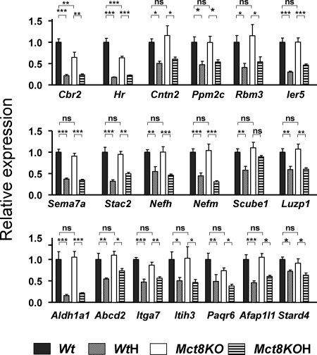
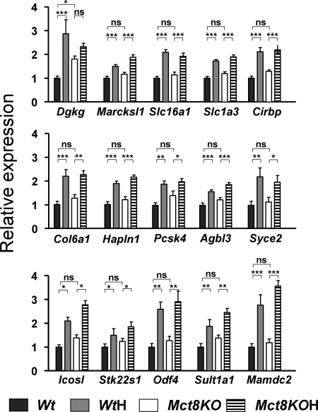
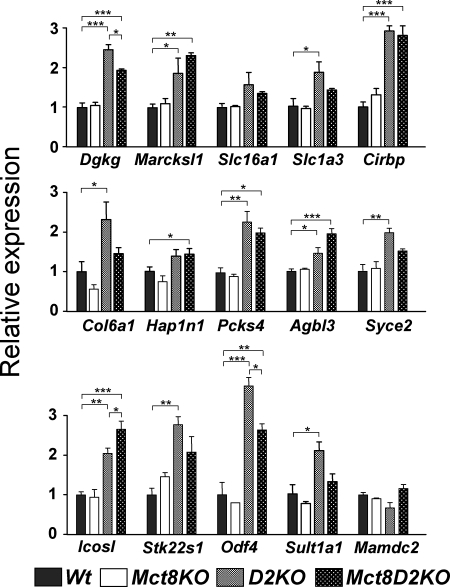
Thyroid hormones influence brain development through the control of gene expression. The concentration of the active hormone T(3) in the brain depends on T(3) transport through the blood-brain barrier, mediated in part by the monocarboxylate transporter 8 (Mct8/MCT8) and the activity of type 2 deiodinase (D2) generating T(3) from T(4). The relative roles of each of these pathways in the regulation of brain gene expression is not known. To shed light on this question, we analyzed thyroid hormone-dependent gene expression in the cerebral cortex of mice with inactivated Mct8 (Slc16a2) and Dio2 genes, alone or in combination. We used 34 target genes identified to be controlled by thyroid hormone in microarray comparisons of cerebral cortex from wild-type control and hypothyroid mice on postnatal d 21. Inactivation of the Mct8 gene (Mct8KO) was without effect on the expression of 31 of these genes. Normal gene expression in the absence of the transporter was mostly due to D2 activity because the combined disruption of Mct8 and Dio2 led to similar effects as hypothyroidism on the expression of 24 genes. Dio2 disruption alone did not affect the expression of positively regulated genes, but, as in hypothyroidism, it increased that of negatively regulated genes. We conclude that gene expression in the Mct8KO cerebral cortex is compensated in part by D2-dependent mechanisms. Intriguingly, positive or negative regulation of genes by thyroid hormone is sensitive to the source of T(3) because Dio2 inactivation selectively affects the expression of negatively regulated genes.


Figures

Figure 1
Figure 1
Effects of Mct8 deficiency and thyroid hormone deprivation on gene expression in the cerebral cortex: positive genes. Gene expression was measured by PCR on TaqMan arrays, using RNAs from control wild-type mice (Wt), hypothyroid Wt mice (WtH), Mct8−/y mice (Mct8KO), and hypothyroid Mct8−/y mice (Mct8KOH) (n = 6 for all groups). Results are expressed as mean ± sem relative to the control Wt value set as 1.0. Significant differences (*, P < 0.05, **, P < 0.01, ***, P < 0.001) between means were determined by two-way ANOVA, the two factors being genotype and thyroid status. Abcd2, ATP-binding cassette, subfamily D (ALD), member 2; Afap1l1, actin filament-associated protein 1-like 1; Aldh1a1, aldehyde dehydrogenase family 1; Cbr2, carbonyl reductase 2; Cntn2, contactin 2 (axonal); Hr, hairless; Ier5, immediate early response 5; Itga7, integrin-α7; Itih3, inter-α-trypsin inhibitor, heavy chain 3; Luzp1, leucine zipper protein 1; Nefh, neurofilament, heavy polypeptide; Nefm, neurofilament, medium polypeptide; Paqr6, progestin and adipoQ receptor family member VI; Ppm2c, protein phosphatase 2C, magnesium-dependent, catalytic subunit; Rbm3, RNA binding motif protein-3; Scube1, signal peptide, CUB domain, EGF-like 1; Sema7a, sema domain, immunoglobulin domain (Ig), and GPI membrane anchor, (semaphorin) 7A; Stac2, SH3 and cysteine-rich domain 2; Stard4, StAR-related lipid transfer (START) domain containing 4.
Figure 2
Figure 2
Effects of Mct8 deficiency and thyroid hormone deprivation on gene expression in the cerebral cortex: negative genes. Gene expression was measured by PCR on TaqMan arrays, using RNAs from control Wt mice, hypothyroid Wt mice (WtH), Mct8−/y mice (Mct8KO), and hypothyroid Mct8−/y mice (Mct8KOH) (n = 6 for all groups). Results are expressed as mean ± sem relative to the control Wt value set as 1.0. Significant differences (*, P < 0.05; **, P < 0.01; ***, P < 0.001) between means were determined by two-way ANOVA, the two factors being genotype and thyroid status. Agbl3, ATP/GTP binding protein-like 3; Cirbp, cold inducible RNA binding protein; Col6a1, collagen, type VI, α1. Dgkg, diacylglycerol kinase, γ; Hapln1, hyaluronan and proteoglycan link protein 1; Icosl, Icos ligand; Mamdc2, MAM domain containing 2; Marcksl1, MARCKS-like 1; Odf4, outer dense fiber of sperm tails 4; Pcsk4, proprotein convertase subtilisin/kexin type 4; Slc1a3, solute carrier family 1 (glial high affinity glutamate transporter), member 3; Slc16a1, solute carrier family 16, member 1 (monocarboxylic acid transporter 1); Stk22s1, serine/threonine kinase 22 substrate 1; Sult1a1, sulfotransferase family, cytosolic, 1A, phenol-preferring, member 1; Syce2, synaptonemal complex central element protein 2.
Figure 3
Figure 3
Effects of D2 deficiency on gene expression in the cerebral cortex: positive genes. Gene expression was measured by PCR on TaqMan arrays (microfluidic cards), using RNAs from control Wt mice, Mct8−/y, Mct8−/yDio2−/− mice (Mct8D2KO), and Dio2−/− mice (D2KO) (n = 3 for all groups). Results are expressed as mean ± sem relative to the control Wt value set as 1.0. #, These samples were lost during the procedure. Significant differences (*, P < 0.05; **, P < 0.01; ***, P < 0.001) between means compared with the control values were determined by one-way ANOVA. All other comparisons for which the significance is not shown were not significant. Gene abbreviations as in Fig. 1.
Figure 4
Figure 4
Effects of D2 deficiency on gene expression in the cerebral cortex: negative genes. Gene expression was measured by PCR on TaqMan arrays (microfluidic cards), using RNAs from control Wt mice, Mct8−/y, Mct8−/yDio2−/− mice (Mct8D2KO), and Dio2−/− mice (D2KO) (n = 3 for all groups). Results are expressed as mean ± sem relative to the control Wt value set as 1.0. Significant differences (*, P < 0.05; **, P < 0.01; ***, P < 0.001) between means compared with the control values were determined by one-way ANOVA. All other comparisons for which the significance is not shown were not significant. Gene abbreviations as in Fig. 2.

Similar articles

Cited by

References

    1. Bernal J 2005 Thyroid hormones and brain development. Vitam Horm 71:95–122 - PubMed
    1. Heuer H, Visser TJ 2009 Minireview: pathophysiological importance of thyroid hormone transporters. Endocrinology 150:1078–1083 - PubMed
    1. St Germain DL, Galton VA, Hernandez A 2009 Minireview: defining the roles of the iodothyronine deiodinases: current concepts and challenges. Endocrinology 150:1097–1107 - PMC - PubMed
    1. Galton VA, Wood ET, St Germain EA, Withrow CA, Aldrich G, St Germain GM, Clark AS, St Germain DL 2007 Thyroid hormone homeostasis and action in the type 2 deiodinase-deficient rodent brain during development. Endocrinology 148:3080–3088 - PubMed
    1. Friesema EC, Ganguly S, Abdalla A, Manning Fox JE, Halestrap AP, Visser TJ 2003 Identification of monocarboxylate transporter 8 as a specific thyroid hormone transporter. J Biol Chem 278:40128–40135 - PubMed

Publication types

MeSH terms