. 2010 May 1;588(Pt 9):1451-68.
doi: 10.1113/jphysiol.2009.186676. Epub 2010 Mar 8.

Regulation of Kv channel expression and neuronal excitability in rat medial nucleus of the trapezoid body maintained in organotypic culture

Affiliations

Regulation of Kv channel expression and neuronal excitability in rat medial nucleus of the trapezoid body maintained in organotypic culture

Huaxia Tong et al. J Physiol. .

Abstract

Principal neurons of the medial nucleus of the trapezoid body (MNTB) express a spectrum of voltage-dependent K(+) conductances mediated by Kv1-Kv4 channels, which shape action potential (AP) firing and regulate intrinsic excitability. Postsynaptic factors influencing expression of Kv channels were explored using organotypic cultures of brainstem prepared from P9-P12 rats and maintained in either low (5 mm, low-K) or high (25 mm, high-K) [K(+)](o) medium. Whole cell patch-clamp recordings were made after 7-28 days in vitro. MNTB neurons cultured in high-K medium maintained a single AP firing phenotype, while low-K cultures had smaller K(+) currents, enhanced excitability and fired multiple APs. The calyx of Held inputs degenerated within 3 days in culture, having lost their major afferent input; this preparation of calyx-free MNTB neurons allowed the effects of postsynaptic depolarisation to be studied with minimal synaptic activity. The depolarization caused by the high-K aCSF only transiently increased spontaneous AP firing (<2 min) and did not measurably increase synaptic activity. Chronic depolarization in high-K cultures raised basal levels of [Ca(2+)](i), increased Kv3 currents and shortened AP half-widths. These events relied on raised [Ca(2+)](i), mediated by influx through voltage-gated calcium channels (VGCCs) and release from intracellular stores, causing an increase in cAMP-response element binding protein (CREB) phosphorylation. Block of VGCCs or of CREB function suppressed Kv3 currents, increased AP duration, and reduced Kv3.3 and c-fos expression. Real-time PCR revealed higher Kv3.3 and Kv1.1 mRNA in high-K compared to low-K cultures, although the increased Kv1.1 mRNA was mediated by a CREB-independent mechanism. We conclude that Kv channel expression and hence the intrinsic membrane properties of MNTB neurons are homeostatically regulated by [Ca(2+)](i)-dependent mechanisms and influenced by sustained depolarization of the resting membrane potential.


Figures

Figure 1
Figure 1. The MNTB maintains projections and neuronal morphology in organotypic culture
A, low magnification microphotograph showing preservation of a transverse brainstem slice after 10 DIV organotypic culture. Dashed line indicates midline; dashed circles indicate locations of the MNTBs. B, DIC image showing a patch pipette recording from an MNTB principle neuron (arrow) in an organotypic slice. C, unilateral dextran rhodamine (dextran) injection in LSO resulted in retrograde labelling in both MNTBs in an organotypic culture (i); higher magnification images show dextran labelled MNTB neurons in high [K+]o (ii) and low [K+]o (iii) cultures. D–F, confocal projection images of MNTB neurons showing co-labelling of dextran (red, D) with Kv3.1b (green, E). Cytoplasmic and putative initial segment region (arrow) staining of Kv3.1b was observed in the merged image (F).
Figure 2
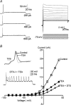
Figure 2. MNTB neurons cultured in high [K+]o (25 mm) medium maintain their phenotypic single AP firing pattern and outward K+ currents
A, left, current-clamp recording shows single fast APs are evoked in response to sustained depolarizing current injection (current magnitude indicated to the right of each trace). Resting membrane potential was adjusted to −70 mV. Right, voltage-clamp shows fast inward sodium currents (*) and sustained outward K+ currents evoked by voltage step commands (10 mV increments; voltage traces below). B, current–voltage (IV) relationships plotted for control (open squares), in the presence of 1 mm TEA (filled squares) and in the presence of both TEA and 100 nm dendrotoxin-I (DTX-I, open diamond). Insets show that TEA broadened the action potential (normalized to the same amplitude) and DTX-I induced multiple action potential firing.
Figure 3
Figure 3. MNTB neurons cultured in low [K+]o (5 mm) medium exhibit reduced K+ currents and increased excitability
A, the mean I–V relations plotted for MNTB neurons from acute brain slices (filled circles), high-K cultures (open squares) and low-K cultures (open triangles). B, the percentage blocks of Kv3 (at +30 mV) and Kv1 (at −30 mV), induced by TEA (1 mm) and DTX-I (100 nm), respectively, are very similar in low (filled bars) and high-K (open bar) cultured neurons. C, a plot of the mean number of APs against the eliciting currents in current-clamp, acute brain slices (filled circles), high-K cultures (open squares) and low-K cultures (open triangles). D, MNTB neurons in low-K cultures (filled bar) exhibited significant (*) lower threshold to elicit APs when compared to neurons from high-K cultures (open bar) and acute brain slices (grey filled bar).
Figure 4
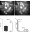
Figure 4. The rise in [Ca2+]i in high-K cultures is mediated by VGCC influx and intracellular store release
A and B, representative fluorescence images of a group of MNTB neurons from organotypic slices with excitation wavelength at 543 nm for dextran tetramethyl-rhodamine (dextran) (A) and 380 nm for Fura2 labelling (B). C, high-K cultures (open bar) displayed significantly greater (*P < 0.05) basal [Ca2+]i to low-K cultures (filled bar). D, summary of percentage increase of [Ca2+]i in high-K cultures during changes of [K+]o from 2.5 mm to 25 mm KCl containing aCSF. Perfusion of VGCC blockers (Nif 10 μm; CTx 2 μm; Aga 200 nm) and inhibition of store release (P < 0.05) by Rya (100 μm) and 2-APB (100 μm) both greatly reduced the change in [Ca2+]i (*P < 0.05).
Figure 5
Figure 5. High [K+]o cultures exhibited greater CREB phosphorylation
Aa, Western blots with the phosphorylated CREB (p-CREB) and total CREB (CREB) antibodies in low-K or high-K cultures from 3 individual experiments. b, representative loading control by α-tubulin shows equal protein loading of one sample onto two lanes labelled with p-CREB (above) and CREB (below). Ba, normalized ratio of phosphorylated CREB (p-CREB) to total CREB is significantly higher in high-K cultures (open bar) than in low-K cultures (filled bar). b, normalized α-tubulin signal ratio showed identical protein loading for lanes labelled with p-CREB and CREB antibody previously in both low (filled bar) and high-K cultures (open bar).
Figure 6
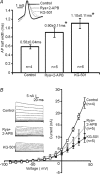
Figure 6. Inhibiting CREB or Ca2+ store release in high [K+]o cultures reduced K+ currents
A, the mean AP half-width increased from 0.58 ± 0.04 ms (control) to 0.90 ± 0.11 ms (Rya+2-APB treated) (*P < 0.05) and to 1.10 ± 0.11 ms (KG-501 treated) (*P < 0.05). Inset: superimposed AP waveforms (normalized to the same amplitude) illustrate broadening of APs with both treatments. B, the average I–V relationships show that Rya and 2-APB incubation reduced K+ currents significantly between +20 and +40 mV (P < 0.05, grey filled squares), and KG-501 treated neurons (filled squares) exhibited less K+ currents between −10 and +40 mV (P < 0.05, filled squares). Insets show a representative current trace for control, Rya+2-APB treated and KG-501 treated neurons; Na+ current was truncated for better focus on K+ currents.
Figure 7
Figure 7. Chronic exposure to VGCC inhibitors attenuated K+ currents in high [K+]o cultures
A, representative responses to depolarizing current steps showed that MNTB neurons raised in high-K (left) failed to maintained the single AP phenotype after incubation with VGCC blockers Nif (nifedipine 10 μm), CTx (ω-conotoxin GVIA 2 μm) and Aga (ω-agatoxin IVA 200 nm). The neuron fired a single AP at a previous subthreshold stimulus (100 pA) followed by multiple APs generated at larger stimuli. Right, the mean AP firing threshold was significantly smaller in treated neurons (*P < 0.05). B, representative traces show the AP waveform was broadened (left) after chronic block of Ca2+ influx through VGCCs. The mean AP half-width increased from 0.49 ± 0.02 ms (control) to 0.66 ± 0.03 ms (treated) (*P < 0.05). C, the mean number of APs plotted against the eliciting currents, showes increased excitability in VGCC blocker treated neurons. D, the average I–V relationship (normalized to the maximum current in control neurons) showes that incubation with VGCC blockers (filled squares) substantially reduced outward K+ currents.
Figure 8
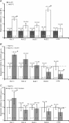
Figure 8. Comparison of K+ channel mRNA expressed in cultures with low [K+]o, high [K+]o and high [K+]o incubated with either CREB antagonist or VGCC blockers
Expression of K+ channel mRNA in the MNTB was estimated by Qrt-PCR. A, expression relative to low-K cultures; high-K cultures showed significantly (*P < 0.05) higher levels of Kv1.1 and Kv3.3 mRNA expression. B, CREB antagonist (KG-501) reduced (*P < 0.05) the mRNA levels of Kv3.3 and c-fos but potentiated Kv1.1 expression in high-K cultures. C, VGCC blockers (nifedipine, ω-conotoxin GVIA and ω-agatoxin IVA) reduced (*P < 0.05) the mRNA levels of Kv1.2, 3.3 and c-fos but potentiated Kv1.1 expression in high-K cultures.
Figure 9
Figure 9. Depolarization-induced homeostatic regulation of K+ channel expression
Upon depolarization, calcium influx through voltage-gated calcium channels (VGCCs) and ligand-gated channels (including NMDA receptors) elevates cytosolic Ca2+ and triggers ryanodine/IP3 sensitive Ca2+ release (CICR) from the endoplasmic reticulum (ER). The amplified Ca2+ signal promotes phosphorylation of CREB, which activates K+ channel transcription, K+ channel expression is up-regulated, and neurons become less excitable with faster action potentials and higher firing threshold.

Comment in

Similar articles

Cited by

References

    1. Barnes-Davies M, Forsythe ID. Pre- and postsynaptic glutamate receptors at a giant excitatory synapse in rat auditory brainstem slices. J Physiol. 1995;488:387–406. - PMC - PubMed
    1. Baxter AW, Wyllie DJ. Phosphatidylinositol 3 kinase activation and AMPA receptor subunit trafficking underlie the potentiation of miniature EPSC amplitudes triggered by the activation of L-type calcium channels. J Neurosci. 2006;26:5456–5469. - PMC - PubMed
    1. Berridge MJ. Neuronal calcium signalling. Neuron. 1998;21:13–26. - PubMed
    1. Best JL, Amezcua CA, Mayr B, Flechner L, Murawsky CM, Emerson B, Zor T, Gardner KH, Montminy M. Identification of small-molecule antagonists that inhibit an activator: coactivator interaction. Proc Natl Acad Sci U S A. 2004;101:17622–17627. - PMC - PubMed
    1. Brew HM, Forsythe ID. Two voltage-dependent K+ conductances with complementary functions in postsynaptic integration at a central auditory synapse. J Neurosci. 1995;15:8011–8022. - PMC - PubMed

Publication types

MeSH terms

LinkOut - more resources