

Structural basis of O6-alkylguanine recognition by a bacterial alkyltransferase-like DNA repair protein
- PMID: 20212037
- PMCID: PMC2859536
- DOI: 10.1074/jbc.M109.093591
Structural basis of O6-alkylguanine recognition by a bacterial alkyltransferase-like DNA repair protein
Abstract
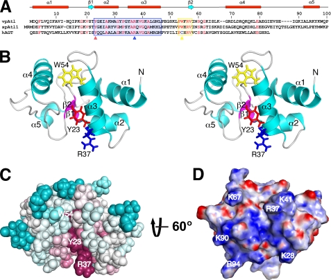
Alkyltransferase-like proteins (ATLs) are a novel class of DNA repair proteins related to O(6)-alkylguanine-DNA alkyltransferases (AGTs) that tightly bind alkylated DNA and shunt the damaged DNA into the nucleotide excision repair pathway. Here, we present the first structure of a bacterial ATL, from Vibrio parahaemolyticus (vpAtl). We demonstrate that vpAtl adopts an AGT-like fold and that the protein is capable of tightly binding to O(6)-methylguanine-containing DNA and disrupting its repair by human AGT, a hallmark of ATLs. Mutation of highly conserved residues Tyr(23) and Arg(37) demonstrate their critical roles in a conserved mechanism of ATL binding to alkylated DNA. NMR relaxation data reveal a role for conformational plasticity in the guanine-lesion recognition cavity. Our results provide further evidence for the conserved role of ATLs in this primordial mechanism of DNA repair.
Figures


Similar articles
-
Flipping of alkylated DNA damage bridges base and nucleotide excision repair.Nature. 2009 Jun 11;459(7248):808-13. doi: 10.1038/nature08076. Nature. 2009. PMID: 19516334 Free PMC article.
-
Alkyltransferase-like protein (Atl1) distinguishes alkylated guanines for DNA repair using cation-π interactions.Proc Natl Acad Sci U S A. 2012 Nov 13;109(46):18755-60. doi: 10.1073/pnas.1209451109. Epub 2012 Oct 29. Proc Natl Acad Sci U S A. 2012. PMID: 23112169 Free PMC article.
-
Alkyltransferase-like protein clusters scan DNA rapidly over long distances and recruit NER to alkyl-DNA lesions.Proc Natl Acad Sci U S A. 2020 Apr 28;117(17):9318-9328. doi: 10.1073/pnas.1916860117. Epub 2020 Apr 9. Proc Natl Acad Sci U S A. 2020. PMID: 32273391 Free PMC article.
-
Repair of O(6)-alkylguanine by alkyltransferases.Mutat Res. 2000 Apr;462(2-3):83-100. doi: 10.1016/s1383-5742(00)00017-x. Mutat Res. 2000. PMID: 10767620 Review.
-
Conserved structural motifs governing the stoichiometric repair of alkylated DNA by O(6)-alkylguanine-DNA alkyltransferase.Mutat Res. 2000 Aug 30;460(3-4):151-63. doi: 10.1016/s0921-8777(00)00024-0. Mutat Res. 2000. PMID: 10946226 Review.
Cited by
-
Atomic-Scale View of Protein-PEG Interactions that Redirect the Thermal Unfolding Pathway of PEGylated Human Galectin-3.Angew Chem Int Ed Engl. 2022 Oct 4;61(40):e202203784. doi: 10.1002/anie.202203784. Epub 2022 Aug 25. Angew Chem Int Ed Engl. 2022. PMID: 35922375 Free PMC article.
-
P53 conformational switching for selectivity may reveal a general solution for specific DNA binding.EMBO J. 2011 Jun 1;30(11):2099-100. doi: 10.1038/emboj.2011.133. EMBO J. 2011. PMID: 21629273 Free PMC article.
-
Multifaceted roles of alkyltransferase and related proteins in DNA repair, DNA damage, resistance to chemotherapy, and research tools.Chem Res Toxicol. 2011 May 16;24(5):618-39. doi: 10.1021/tx200031q. Epub 2011 Apr 28. Chem Res Toxicol. 2011. PMID: 21466232 Free PMC article. Review.
-
The 100-protein NMR spectra dataset: A resource for biomolecular NMR data analysis.Sci Data. 2024 Jan 4;11(1):30. doi: 10.1038/s41597-023-02879-5. Sci Data. 2024. PMID: 38177162 Free PMC article.
-
Understanding the importance of low-molecular weight (ethylene oxide- and propylene oxide-induced) DNA adducts and mutations in risk assessment: Insights from 15 years of research and collaborative discussions.Environ Mol Mutagen. 2019 Mar;60(2):100-121. doi: 10.1002/em.22248. Epub 2018 Dec 10. Environ Mol Mutagen. 2019. PMID: 30536466 Free PMC article. Review.
References
-
- Verbeek B., Southgate T. D., Gilham D. E., Margison G. P. (2008) Br. Med. Bull. 85, 17–33 - PubMed
-
- Daniels D. S., Woo T. T., Luu K. X., Noll D. M., Clarke N. D., Pegg A. E., Tainer J. A. (2004) Nat. Struct. Mol. Biol. 11, 714–720 - PubMed
-
- Duguid E. M., Rice P. A., He C. (2005) J. Mol. Biol. 350, 657–666 - PubMed
-
- Margison G. P., Butt A., Pearson S. J., Wharton S., Watson A. J., Marriott A., Caetano C. M., Hollins J. J., Rukazenkova N., Begum G., Santibáñez-Koref M. F. (2007) DNA Repair 6, 1222–1228 - PubMed
Publication types
MeSH terms
Substances
Grants and funding
LinkOut - more resources
Full Text Sources
Miscellaneous
