. 2010 May 14;285(20):15076-15087.
doi: 10.1074/jbc.M109.078915. Epub 2010 Mar 8.

Mammalian Ste20-like kinase (Mst2) indirectly supports Raf-1/ERK pathway activity via maintenance of protein phosphatase-2A catalytic subunit levels and consequent suppression of inhibitory Raf-1 phosphorylation

Affiliations

Mammalian Ste20-like kinase (Mst2) indirectly supports Raf-1/ERK pathway activity via maintenance of protein phosphatase-2A catalytic subunit levels and consequent suppression of inhibitory Raf-1 phosphorylation

Geoffrey K Kilili et al. J Biol Chem. .

Abstract

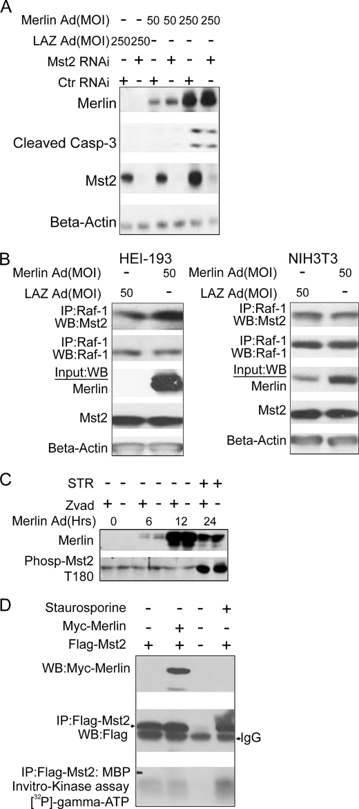
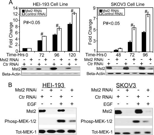
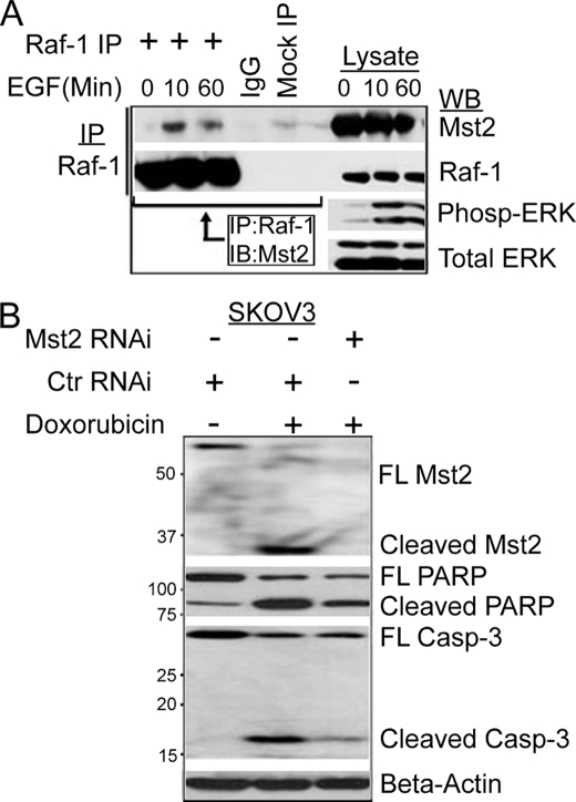
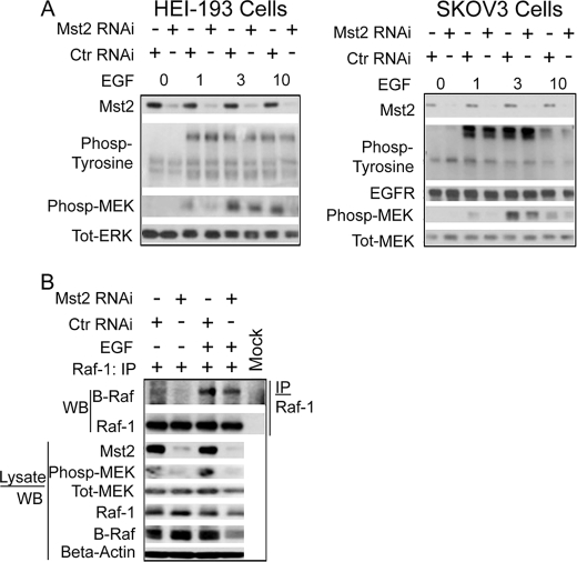
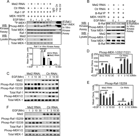
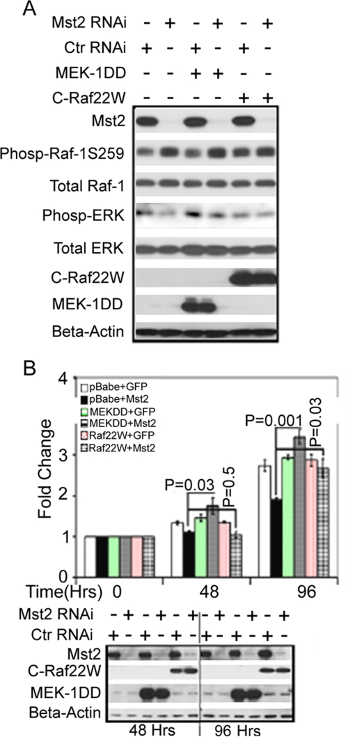
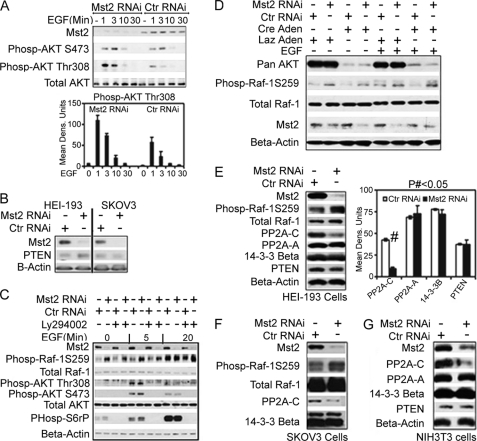
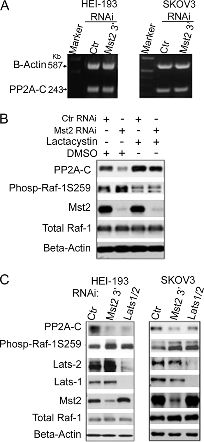
Many tumor suppressor proteins act to blunt the effects of mitogenic signaling pathways. Loss of function mutations in the merlin tumor suppressor underlie neurofibromatosis type 2 (NF2), a familial autosomal dominant cancer syndrome. Studies of Drosophila suggest that Hippo (hpo) is required for inhibition of cell proliferation mediated by dMer, the orthologue of human merlin. Mammalian sterile 20-like kinase-2 (Mst2) is a mammalian Hpo orthologue, and numerous studies implicate Mst2 as a tumor suppressor. Mst2 is negatively regulated by the proto-oncoprotein Raf-1 in a manner independent of the kinase activity of Raf-1. We sought to determine whether, in mammalian cells, merlin could positively regulate Mst2. We also sought to determine whether Mst2, in addition to being negatively regulated by Raf-1, might itself reciprocally regulate Raf-1. In contrast to findings from Drosophila, we find no evidence that mammalian merlin positively regulates mammalian Mst2. Instead, surprisingly, RNA interference silencing of Mst2 leads to elevated inhibitory phosphorylation of Raf-1 at Ser-259 and impaired Raf-1 kinase activity. Consequent to this, ERK pathway activation and cell proliferation are attenuated. Phosphatase-2A (PP2A) dephosphorylates Raf-1 Ser-259 in response to mitogens. Interestingly RNA interference silencing of Mst2 triggers a striking proteasome-dependent decrease in the levels of the catalytic subunit of PP2A (PP2A-C). A similar effect is achieved upon silencing of large tumor suppressor (LATS)-1 and LATS2, direct substrates of Mst2. Our studies reveal a more complex role for Mst2 than previously thought. The Mst2 --> LATS1/2 pathway, by maintaining PP2A-C levels, may, in some situations, positively affect mitogenic signaling.


Figures

FIGURE 1.
FIGURE 1.
Merlin does not function upstream of or positively regulate MST2. A, knockdown of Mst2 does not inhibit merlin-meditated cell death. HEI-193 cells were transfected with either human Mst2-specific RNAi or control (Ctr) (mouse-specific Mst2 RNAi) and infected with a LacZ (LAZ) or NF2-overexpressing adenovirus (Ad) at a 50 or 250 multiplicity of infection (MOI) 24 h later. Cells were lysed in RIPA buffer 48 h after infection and subjected to immunoblotting with the indicated antibodies. B, merlin does not promote disruption of the Raf-1-Mst2 complex. co-immunoprecipitations (IP) in HEI-193 cells (left) and NIH3T3 cells (right). Cells were lysed in Lysis buffer A, and 500 μg of total lysate was used. WB, Western blot. C, merlin does not promote phosphorylation of Mst2 on Thr-180. Total RIPA buffer lysates from HEI-193 cells infected with NF2-overexpressing adenovirus for the indicated amount of time were subjected to immunoblot analysis with phospho-Mst2 (Phosp-Mst2) Thr-180 (T180) antibody. STR = staurosporine D, merlin overexpression does not affect Mst2 kinase activity. 0.3 μg of FLAG-Mst2 was co-transfected with 1 μg of Myc-merlin into HEK293 cells, and 18 h later, cells were lysed, and 500 μg of total lysate was subjected to immunoprecipitation with FLAG antibody. 50% of immunoprecipitated beads-FLAG-Mst2 complex was used in an in vitro kinase assay with myelin basic protein as substrate.
FIGURE 2.
FIGURE 2.
Mst2 is required for optimal activation of the Ras/Raf/MEK/ERK mitogen-activated pathway and cell proliferation. A, RNAi silencing of Mst2 reduces cell proliferation. Left, HEI-193; right, SKOV3. Cells were transfected with 100 nm human-specific Mst2 or control (Ctr) (mouse) RNAi. 24 h later, cells were serum-starved overnight and trypsinized, and equal numbers were plated in triplicate per condition. At the indicted time points, cells were counted with a hemocytometer. Parallel samples were lysed at each time point for immunoblot analysis with the indicated antibodies. Numbers represent -fold change ± S.E. relative to T0. (# indicates p < 0.05, n = 3. Control values (time 0) were set to 1 and not subjected to statistical analysis. B, Mst2 RNAi impairs MAPK signaling in response to EGF stimulation. HEI-193 cells (left) and SKOV3 cells (right) were treated with human-specific Mst2 RNAi or control (mouse) RNAi (100 nm). After 48 h, cells were serum-starved for 18–20 h and then treated with 50 ng/ml EGF (+EGF) or vehicle (−EGF) for 10 min. Lysates were subjected to immunoblotting with the indicated antibodies. Phosp-MEK-1/2, phospho-MEK1/2; Tot-Mek-1, total Mek1.
FIGURE 3.
FIGURE 3.
Mitogens enhance assembly of the Raf-1-Mst2 complex; Mst2 is required for optimal doxorubicin-induced cell death. A, EGF promotes assembly of the Raf-1-Mst2 complex. NIH3T3 cells were serum-starved overnight and then treated with 50 ng/ml EGF for the indicated time points before lysis and immunoprecipitation (IP) with Raf-1 antibody. Immunoprecipitates were then immunoblotted (IB) with anti-Mst2 antibody. Phospho-ERK (Pho-ERK) immunoblotting was used as a positive control readout for activation of the Ras-Raf/ERK pathway by EGF. WB, Western blot. Tot ERK, total ERK. B, in response to genotoxic stress, knockdown of Mst2 promotes cell survival. SKOV3 or HEI-193 cells were transfected with human-specific Mst2 RNAi or control RNAi (Ctr RNAi, mouse). 48 h later, cells were treated with either 10 μm doxorubicin or vehicle (DMSO) for 8 h and then lysed. Equal amounts were immunoblotted with antibodies to the indicated apoptotic markers. FL, full length. PARP, poly(ADP-ribose) polymerase; casp-3, caspase-3.
FIGURE 4.
FIGURE 4.
Mst2 is not required for optimal EGF receptor activation or for the integrity of the Raf-1-B-Raf complex. A, Mst2 RNAi does not affect EGF receptor phosphorylation. Left, HEI-193; Right, SKOV3. (Please note that these are the same total lysates saved from the samples used to immunoprecipitate Raf-1 for the kinase assay in Fig. 5A, also used for supplemental Fig. 5A; hence the phospho-MEK (phosp-MEK), total MEK (Tot-MEK), and Mst2 blots have been reproduced in these figures for the convenience of the reader). Cells were treated as indicated in Fig. 5A below and subjected to immunoblot analysis with the indicated antibodies. Total ERK (Tot-ERK) immunoblotting was performed as a gel loading control. Ctr RNAi, control RNAi. B, integrity of the Raf-1-B-Raf complex is not affected by Mst2 RNAi. HEI-193 cells were grown and transfected with 100 nm human-specific Mst2 or control (mouse) RNAi. After 48 h cells, were serum-starved for 18–20 h and then treated with EGF or vehicle for 10 min. Raf-1 was then immunoprecipitated (IP) and subjected to SDS-PAGE and immunoblot analysis with the indicated antibodies. WB, Western blot.
FIGURE 5.
FIGURE 5.
Mst2 positively regulates Raf-1 activation via suppression of the inhibitory phosphorylation of Raf-1 on Ser-259; Mst2 is not required for B-Raf activation. A, Raf-1 kinase activity is impaired upon Mst2 RNAi. SKOV3 cells were grown and transfected with 100 nm human-specific Mst2 RNAi or control (Ctr) (mouse) RNAi. After 48 h, cells were serum-starved for 18–20 h and then treated with EGF for the indicated time points before lysis in Lysis buffer A. Raf-1 was then immunoprecipitated (IP) and assayed using purified MEK1 K97R as a substrate. A part of the total lysate was saved and separately subjected to immunoblotting with phospho-MEK1/2 Ser-217/221, total MEK, or Mst2 antibodies. (Note: These are the same lysates used in Fig. 4A (SKOV3 blot) and supplemental Fig. 5A; hence the phospho-MEK (Phosp-MEK-1), total MEK, and Mst2 blots have been reproduced for the convenience of the reader.) For the in vitro kinase assay, MEK phosphorylation was quantitated with ImageJ as shown in the bar graphs. Data shown are means ± S.E. Replicates were analyzed by Student's t test (n = 3, # indicates p = 0.02, Dens. Units indicates densitometric units). WB, Western blot. B, the kinase activity of B-Raf is not affected by Mst2 knockdown. Cell handling, B-Raf immunoprecipitation, and assaying were performed as for Raf-1 in A above. C, Ser-259, an inhibitory phosphoacceptor site on Raf-1, is hyperphosphorylated upon silencing of Mst2. HEI-193 cells were treated either with 100 nm Mst2 targeting RNAi (human Mst2-RNAi) or with control RNAi (mouse). 48 h later, cells were serum-starved and then stimulated with 50 ng/ml EGF for the indicated time points before lysis. Lysates were immunoblotted with the indicated antibodies. D and E, densitometric analysis of the effect of Mst2 RNAi on Raf-1 Ser-259 phosphorylation and MEK1/2 217/221 phosphorylation. The experiment in C was repeated three times, and phospho-MEK1/2 Ser-217/221 (D) and phospho-Raf-1 Ser-259 (E) were quantified with ImageJ software. Each data point represents the average of three independent experimental densitometry values of that particular time point ± S.E. Data were analyzed by Student's t test. # indicates p < 0.05 for control versus Mst2 RNAi samples indicated. F, similar results of hyperphosphorylation of Raf-1 Ser-259 upon Mst2 silencing were obtained using a second human-specific Mst2 oligonucleotide targeting a different part of the Mst2 transcript.
FIGURE 6.
FIGURE 6.
Ectopic, stable expression of either constitutively active MEK1 or constitutively active Raf-1 rescues the proliferation defect in Mst2 knockdown Cells. A, expression of MEK1DD (MEK1-S218D/S221D) or C-Raf 22W (a C-terminal construct expressing amino acids 321–552 and thus missing Ser-259) has no effect on the elevation in endogenous Raf-1 Ser-259 phosphorylation (Phosp-Raf-1S259) incurred upon silencing of Mst2 but can partially reverse the resulting decrease in ERK phosphorylation in cells maintained in 10% serum. HEI-193 cells stably overexpressing MEK1DD, C-Raf 22W, or empty pBabe-puro vector were treated with human-specific Mst2 RNAi (oligo-1) or control RNAi (Ctr RNAi) (GFP). 72 h later, cell extracts were prepared and immunoblotted with the indicated antibodies. B, constitutively active MEK-DD or Raf-22W can rescue HEI-193 cells from the reduction in cell proliferation incurred upon Mst2 silencing. Top panel, the stable HEI-193-pBabe-puro, MEK1DD, or C-Raf 22W cell lines were treated with either control RNAi (+GFP) or Mst2 RNAi (+Mst2) as described in A above. A cell proliferation assay was performed as described in the legend for Fig. 2A. An unpaired Student's t test was performed to ascertain that overexpression of MEK1DD or C-Raf 22W resulted in significant rescue of cell proliferation upon Mst2 knockdown relative to the pBabe-puro Mst2 knockdown cell line (control cell line). The bottom panel documents the level of Mst2 RNAi and MEK-DD or Raf-22W expression during the time course of the study. Data shown are means ± S.E.
FIGURE 7.
FIGURE 7.
Mst2 functions to maintain levels of the catalytic subunit of PP2A (PP2A-C); although silencing of Mst2 enhances Akt activation, Akt is not required for phosphorylation of Raf-1 at Ser-259. A, Mst2 RNAi leads to hyperactivation of the PI3K/Akt pathway. HEI-193 cell RNAi transfection, handling, cell lysis, and immunoblotting were performed as described in the legend for Fig. 5A. Ctr RNAi, control RNAi; Phosp-AKT S473, phospho-AKT Ser-473. Data shown are means ± S.E. B, PTEN levels are not affected by silencing of Mst2 in both HEI-193 and SKOV3 cells. C and D, neither PI3K inhibitor LY294002 (C) nor deletion of all three Akt isoforms (D) could affect the Raf-1 Ser-259 hyperphosphorylation observed upon silencing Mst2. For C, HEI-193 cells were treated with vehicle (−) (DMSO) or 50 μm LY294002 (+) as indicated. Cells were then treated with EGF for the indicated time points. Equal amounts of total lysates were then separated by SDS-PAGE and immunoblotted with the indicated antibodies. For D, akt2/3−/−;akt-1flox/flox MEFs were treated with Cre recombinase-expressing adenovirus (Cre Aden) to obtain Akt MEFs expressing no Akt isoform. Control MEFs were treated with Lac-Z (Laz in the figure)-expressing adenovirus. MEFs were then transfected with mouse-specific Mst2 RNAi. Human Mst2 RNAi was used as control. After 48 h, MEFs were treated with EGF or vehicle (0) as indicated. MEFs were then lysed in RIPA buffer, and equal amounts of lysate were subjected to SDS-PAGE and immunoblot analysis with the indicated antibodies. E–G, levels of PP2A-C are diminished upon Mst2 RNAi. HEI-193 (E) or SKOV3 (G) cells were treated with human-specific Mst2 or control RNAi (mouse). NIH3T3 cells (F) were treated with the different mouse-specific oligo set described under “Experimental Procedures” and the human Mst2 RNAi (oligo-1) as control. Lysates were then subjected to immunoblot analysis for the expression levels of PP2A-C, PP2A-A, 14-3-3-β, or PTEN. Only the expression levels of PP2A-C were found to be significantly affected by Mst2 silencing. E, in the bar graph, # indicates p < 0.001 for control versus Mst2 RNAi samples. Data shown are means ± S.E.
FIGURE 8.
FIGURE 8.
Mst2 stabilizes PP2A-C through a post-translational mechanism involving stabilization of the PP2A polypeptide. PP2A-C stabilization requires the Mst2 substrate kinases LATS1/2. A, Mst2 RNAi has no effect on PP2A-C transcript levels. HEI-193 or SKOV3 cells were subjected to Mst2 RNAi targeting the 3′-untranslated region of human Mst2 transcript (the RNA samples for this study were prepared in parallel with cells used in panel C, which shows the efficacy of Mst2 RNAi). For control (Ctr), GFP-specific RNAi was used. RNA was prepared and subjected to reverse transcription-PCR to assess PP2A-C levels. β-Actin reverse transcription-PCR served as a loading control. B, the proteasome inhibitor lactacystin restores PP2A-C levels depleted upon Mst2 RNAi, coincident with a decrease in Raf-1 Ser-259 phosphorylation. HEI-193 cells were treated with human-specific Mst2 RNAi (oligo-1) and 10 μm lactacystin 48 h later for 18 h as indicated. GFP RNAi was used as control. Extracts were prepared and immunoblotted with the indicated antibodies. C, RNAi silencing of LATS1/2 results in destabilization of PP2A-C and coincident increased Raf-1 Ser-259 phosphorylation. As indicated, either HEI-193 or SKOV3 cells were subjected to the 3′-untranslated region targeting the Mst2 RNAi oligo described in A above, which is different from the Mst2 oligo-1 used in Fig. 7, E and F, or the murine oligo set used for NIH3T3 cells (Fig. 7G). A sample, contemporaneously treated with human-specific LATS1 and LATS2 RNAi oligonucleotides, was also included. GFP RNAi was used as control. Cell extracts were prepared and subjected to immunoblotting with the indicated antibodies. Phos-Raf-1S259, phosho-Raf-1 Ser-259.

Similar articles

Cited by

References

    1. Brown M. D., Sacks D. B. (2009) Cell. Signal. 21, 462–469 - PMC - PubMed
    1. Widmann C., Gibson S., Jarpe M. B., Johnson G. L. (1999) Physiol. Rev. 79, 143–180 - PubMed
    1. Lowy D. R., Willumsen B. M. (1993) Annu. Rev. Biochem. 62, 851–891 - PubMed
    1. Dhillon A. S., Pollock C., Steen H., Shaw P. E., Mischak H., Kolch W. (2002) Mol. Cell. Biol. 22, 3237–3246 - PMC - PubMed
    1. Zimmermann S., Moelling K. (1999) Science 286, 1741–1744 - PubMed

Publication types