. 2010 Mar 15;207(3):579-89.
doi: 10.1084/jem.20090516. Epub 2010 Mar 8.

Myeloid DAP12-associating lectin (MDL)-1 regulates synovial inflammation and bone erosion associated with autoimmune arthritis

Affiliations

Myeloid DAP12-associating lectin (MDL)-1 regulates synovial inflammation and bone erosion associated with autoimmune arthritis

Barbara Joyce-Shaikh et al. J Exp Med. .

Abstract

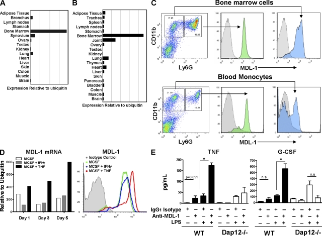
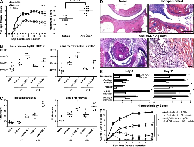
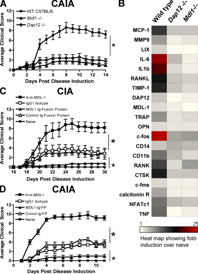
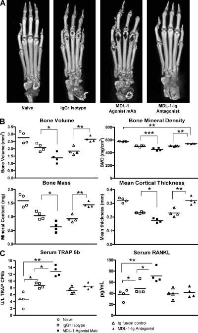
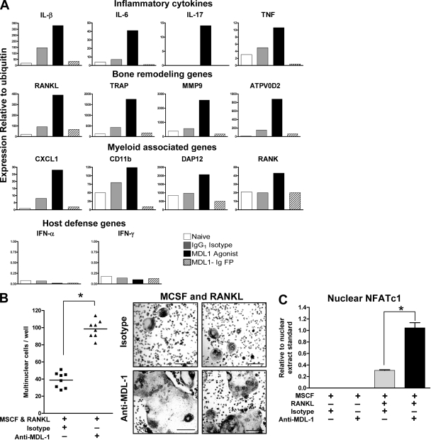
DNAX adaptor protein 12 (DAP12) is a trans-membrane adaptor molecule that transduces activating signals in NK and myeloid cells. Absence of functional Dap12 results in osteoclast defects and bone abnormalities. Because DAP12 has no extracelluar binding domains, it must pair with cell surface receptors for signal transduction. There are at least 15 known DAP12-associating cell surface receptors with distinct temporal and cell type-specific expression patterns. Our aim was to determine which receptors may be important in DAP12-associated bone pathologies. Here, we identify myeloid DAP12-associating lectin (MDL)-1 receptor (also known as CLEC5A) as a key regulator of synovial injury and bone erosion during autoimmune joint inflammation. Activation of MDL-1 leads to enhanced recruitment of inflammatory macrophages and neutrophils to the joint and promotes bone erosion. Functional blockade of MDL-1 receptor via Mdl1 deletion or treatment with MDL-1-Ig fusion protein reduces the clinical signs of autoimmune joint inflammation. These findings suggest that MDL-1 receptor may be a therapeutic target for treatment of immune-mediated skeletal disorders.


Figures

Figure 1.
Figure 1.
Regulation of MDL-1 receptor expression. (A and B) Bone marrow cells and joint tissues show the highest level of MDL-1 expression. Quantitative PCR analysis of human (A) and mouse (B) anatomy panels generated from pooled tissue samples from at least five donors. (C) MDL-1 is expressed on murine granulocytes (CD11b + Ly6G high) and monocytes (CD11b + Ly6G low) from bone marrow and peripheral blood. Histograms in gray color are rIgG2a isotype staining controls. Data are representative of three experiments. (D) TNF but not IFN-γ promotes MDL-1 expression. Murine bone marrow cells were cultured with MCSF in the presence of IFN-γ or TNF and mRNA was isolated for MDL-1 expression analysis by Q-PCR. Cell surface expression of MDL-1 was determined by flow cytometry. Data representative of at least three separate experiments with similar results. (E) Anti–MDL-1 activates bone marrow–derived macrophages to produce TNF and G-CSF. Bone marrow cells isolated from three mice were cultured with indicated antibodies and/or LPS for 36 h. Culture supernatants were assayed for secreted cytokines by Luminex assay. * indicates significance (P < 0.01) as determined by Student’s t test. Results are representative of three separate experiments with similar results.
Figure 2.
Figure 2.
MDL-1 activation enhances autoimmune arthritis. (A) Treatment with anti–MDL-1 agonist mAb exacerbates CAIA. B10RIII mice (n = 5/group) were given arthrogen to induce arthritis. Mice treated with anti–MDL-1 mAb (clone DX163) at the time of arthrogen treatment showed disease exacerbation compared with isotype controls. Maximum clinical score of individual mice from two separate anti–MDL-1 treatment studies is shown on the right. Results are representative of at least four experiments. (B) Anti–MDL-1 treatment increases the absolute number of bone marrow granulocytes and monocytes. Each data point is FACS analysis of cells extracted from two tibias. (C) Peripheral blood peroxidase-positive neutrophils and macrophages are elevated in anti–MDL-1 treated mice, as shown by ADVIA analysis. The studies in B and C were performed twice. (D) Representative H&E-stained micrographs of metatarsal-phalange joints from the study shown in A. Bars: (top left, bottom left, and top right) 200 µm; (bottom right) 60 µm. The anti–MDL-1 agonist treatment group showed intense neutrophil and macrophage infiltration, as well as pannus tissue formation with extensive bone erosion. (E) Histopathology was performed in a masked fashion. Leukocyte infiltration and percentage of PMN infiltration were determined in the synovium and joint space. Percent PMNs: 1 = <20%, 2 = 20–40%, 3 = 40–60%, and 4 = >60%. Pannus tissue formation, cartilage destruction, and cortical bone erosions were assessed as described in the Materials and methods section. The severity was graded on a scale of 0–4. Comparisons between anti–MDL-1 and isotype control were determined using the Mann Whitney U test. * indicates P < 0.05 and is considered statistically significant. (F) Depletion of granulocytes/monocytes reduced anti–MDL-1–driven CAIA. Date shown are summary of two separate experiments (n = 10 per treatment group). Disease was induced as in A with additional groups that were pretreated on day −1 with anti-GR1 mAb (clone RB6-8C5, rIgG2a isotype) that depletes Ly6G+ and a subset of Ly6C+ cells. Disease was induced on day 0 and mice were given anti–MDL-1 mAb (clone DX163) or an IgG1 isotype control. Depletion of GR1+ populations was confirmed by flow cytometry (Fig. S4).
Figure 3.
Figure 3.
MDL-1 blockade inhibits autoimmune arthritis. (A) C57BL/6 control, Dap12−/−, and Mdl1−/− mice were injected with arthrogenic mAbs to induce CAIA. Mean clinical scores (n = 5/group) ± SEM are shown. Result is representative of two studies. * indicates statistical significance. (B) Hind paw mRNA from A was isolated for gene expression analysis. The absolute values of the inflammatory genes are shown in Fig. S7. (C) MDL-1 activation enhanced T cell– and myeloid cell–dependent arthritis. B10RIII mice were immunized with bovine type II collagen emulsified in CFA at day 0 to induce collagen-induced arthritis. Anti–MDL-1 agonist (clone DX163) was given on day 18 of immunization. Statistical significance was determined by analysis of variance (ANOVA). Results are representative of two studies. (D) B10RIII mice (n > 5/group) were given 1.5 mg of Arthrogen to induce CAIA. MDL-1 Ig fusion protein or control proteins were administrated at the time of Arthrogen treatment. * indicates statistical significance (P < 0.001) as determined by ANOVA analysis.
Figure 4.
Figure 4.
MDL-1 blockade inhibits bone erosion and inflammation. (A) Hind paws from mice in Fig. 3D were harvested on day 11 and prepared for MicroCT analysis. Images show high resolution three-dimensional rendering of MicroCT scans. MDL-1 Ig-fusion protein (antagonist)–treated mice show no sign of cortical bone destruction, whereas the agonist MDL-1 antibody (clone: DX163) treated mice show greater bone destruction than control IgG1-treated mice. Results are representative of three separate experiments. (B) Quantification of bone integrity indicates increased bone loss in anti–MDL-1 agonist-treated mice. Comparable regions of interest (ROI) consisting of three metatarsal joints from each mouse were selected for analysis. Bone volume (BV), bone mass (BM), bone mineral density (BMD = BM/BV mg/cm3), and mean cortical thickness (mm) were quantified from MicroCT scans using GE MicroView software v2.2. Student’s t test was used to determine significance where ***, P < 0.001; **, P < 0.01; *, P < 0.05. (C) ELISA quantification of Serum TRAP 5b and RANKL. Day 20 serum levels of TRAP5b and RANKL from individual mice are shown. TRAP5b levels from day 11 also show similar results. * indicates P < 0.05 by Student’s t test analysis.
Figure 5.
Figure 5.
MDL-1 activation promotes NFATc1 nuclear translocation and enhances osteoclast formation. (A) Gene expression of pooled (n = 3) hind paws treated as in Fig. 3D were harvested at day 4 and prepared for Q-PCR analysis. Data are representative of four experiments. (B) Bone marrow cells were cultured in plastic dishes in the presence of 10 ng/ml RANKL and MCSF. Anti–MDL-1, but not isotype control (or medium control; not depicted) increased the number multinuclear cells (left) and osteoclast size (right). Data are representative of at least four separate experiments. Bars, 200 µm. (C) MDL-1 regulates NFATc1 nuclear expression. C57BL/6 bone marrow cells were cultured with indicated growth factors and antibodies. Cells were cultured for 6 d and nuclear lysates prepared for Active Motif Nuclear NFATc1 colorimetric assay. Nuclear NFATc1 was detected by a plate-bound NFAT oligonucleotide consensus sequence (5′-AGGAAA-3′), a primary NFATc1-specific antibody, and a secondary HRP-conjugated mAb. Results are representative of two studies. * indicates significance (P < 0.01) as determine by ANOVA analysis.

Similar articles

Cited by

References

    1. Aoki N., Zganiacz A., Margetts P., Xing Z. 2004. Differential regulation of DAP12 and molecules associated with DAP12 during host responses to mycobacterial infection. Infect. Immun. 72:2477–2483 10.1128/IAI.72.5.2477-2483.2004 - DOI - PMC - PubMed
    1. Bakker A.B., Baker E., Sutherland G.R., Phillips J.H., Lanier L.L. 1999. Myeloid DAP12-associating lectin (MDL)-1 is a cell surface receptor involved in the activation of myeloid cells. Proc. Natl. Acad. Sci. USA. 96:9792–9796 10.1073/pnas.96.17.9792 - DOI - PMC - PubMed
    1. Barck K.H., Lee W.P., Diehl L.J., Ross J., Gribling P., Zhang Y., Nguyen K., van Bruggen N., Hurst S., Carano R.A. 2004. Quantification of cortical bone loss and repair for therapeutic evaluation in collagen-induced arthritis, by micro-computed tomography and automated image analysis. Arthritis Rheum. 50:3377–3386 10.1002/art.20557 - DOI - PubMed
    1. Chen S.T., Lin Y.L., Huang M.T., Wu M.F., Cheng S.C., Lei H.Y., Lee C.K., Chiou T.W., Wong C.H., Hsieh S.L. 2008. CLEC5A is critical for dengue-virus-induced lethal disease. Nature. 453:672–676 10.1038/nature07013 - DOI - PubMed
    1. Henriksen K., Tanko L.B., Qvist P., Delmas P.D., Christiansen C., Karsdal M.A. 2007. Assessment of osteoclast number and function: application in the development of new and improved treatment modalities for bone diseases. Osteoporos. Int. 18:681–685 10.1007/s00198-006-0286-8 - DOI - PubMed

Publication types

MeSH terms