. 2010 Mar 15;207(3):475-89.
doi: 10.1084/jem.20090831. Epub 2010 Mar 8.

microRNA-29a induces aberrant self-renewal capacity in hematopoietic progenitors, biased myeloid development, and acute myeloid leukemia

Affiliations

microRNA-29a induces aberrant self-renewal capacity in hematopoietic progenitors, biased myeloid development, and acute myeloid leukemia

Yoon-Chi Han et al. J Exp Med. .

Abstract

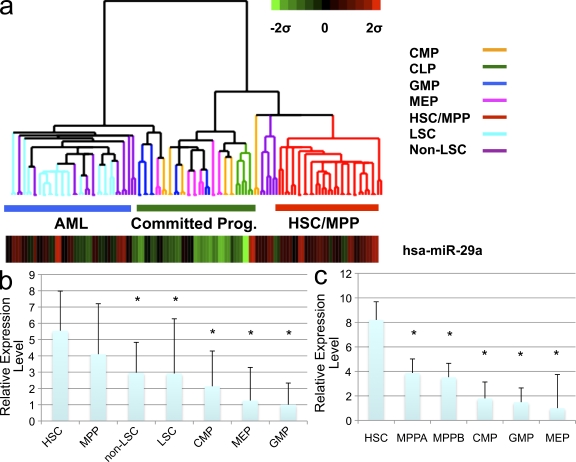
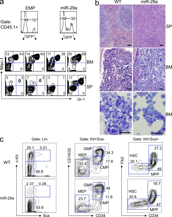
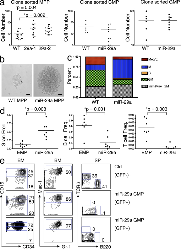
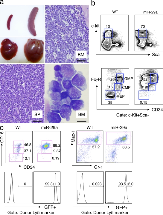
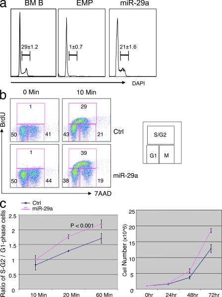
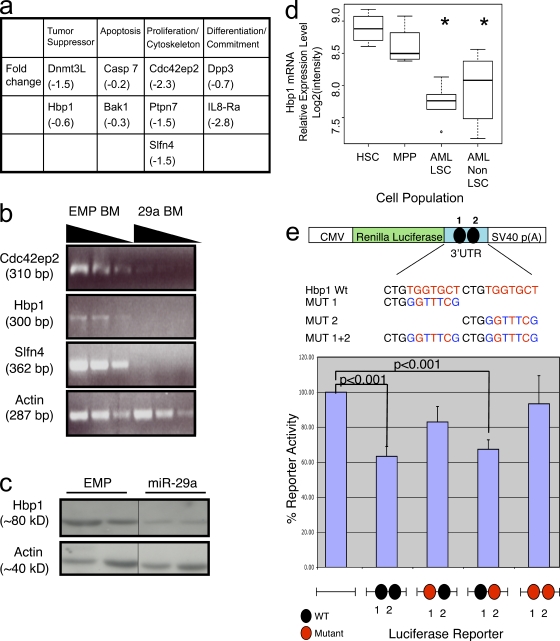
The function of microRNAs (miRNAs) in hematopoietic stem cells (HSCs), committed progenitors, and leukemia stem cells (LSCs) is poorly understood. We show that miR-29a is highly expressed in HSC and down-regulated in hematopoietic progenitors. Ectopic expression of miR-29a in mouse HSC/progenitors results in acquisition of self-renewal capacity by myeloid progenitors, biased myeloid differentiation, and the development of a myeloproliferative disorder that progresses to acute myeloid leukemia (AML). miR-29a promotes progenitor proliferation by expediting G1 to S/G2 cell cycle transitions. miR-29a is overexpressed in human AML and, like human LSC, miR-29a-expressing myeloid progenitors serially transplant AML. Our data indicate that miR-29a regulates early hematopoiesis and suggest that miR-29a initiates AML by converting myeloid progenitors into self-renewing LSC.


Figures

Figure 1.
Figure 1.
miR-29a is expressed at high levels in human HSCs as well as human AML. (a) Heat map of miR-29a expression. Expression was normalized against sno-R2 and data are presented as a z-score. Lineage committed progenitors (committed prog.) include CMP, GMP, MEP, and CLP, which were sorted as described in the text. (b) Relative normalized expression levels of miR-29a in FACS-sorted normal human BM populations and human AML shows that miR-29a is expressed at highest levels in HSC. (c) Relative normalized expression levels of miR-29a expression in FACS-purified mouse HSC and lineage committed progenitors shows a dramatic decrease in miR-29a expression in MPPs (MPPA, LinKit+Sca+CD34+Flk2; MPPB, LinKit+Sca+CD34+Flk2+). Asterisks signify statistically significant differential expression of indicated population compared with HSC (q < 0.05). Error bars indicate SD from at least five independent samples.
Figure 2.
Figure 2.
Ectopic expression of miR-29a in mouse HSC/progenitors induces myeloproliferative disease in primary chimeras. (a) Flow cytometric analysis of the BM and spleen of miR-29a–transduced or empty virus control (EMP) long-term engrafted (3–5 mo) primary chimeras reveals that miR-29a induces monocytic/granulocytic hyperplasia. (b) Primary miR-29a chimeric mice exhibit signs of myeloproliferative disease, including increased splenic extramedullary hematopoiesis, as well as granulocytic and megakaryocytic hyperplasia in both the spleen and BM. Bars, 50 µm. Cytospin preparations of the BM were Wright-Giemsa stained to reveal a predominance of maturing granulocytic precursors and near-absence of erythroid precursors in miR-29a mice relative to normal controls (bottom). Bars, 50 µm. (c) The myeloproliferative phenotype is associated with changes in the immature hematopoietic compartment, manifested by increased proportions of LinKit cells and phenotypic HSC, as well as the relative expansion of normal myeloid progenitor populations. These findings are representative of at least four independent experiments with at least 10 mice in the EMP and miR-29a–transduced groups.
Figure 3.
Figure 3.
miR-29a expression alters the proliferation, differentiation, and self-renewal capacity of hematopoietic progenitors. (a) Clone-sorted miR-29a MPPs (sorted either as LinKit+Sca+CD34+FLK2 or LinKit+Sca+CD34+SLAM) show a higher proliferative capacity in liquid culture compared with WT MPP, but this difference is not observed in CMP or GMP. 29a-1 and 29a-2 represent different miR-29a primary chimeric mice. (b) Photomicrographs of representative colonies formed by cloned sorted WT MPP or miR-29a MPP. Bars, 50 µm. (c) Lineage potential was assessed based on evaluation of cytospin preparations from clone-sorted MPP in vitro liquid cultures, demonstrating that miR-29a promotes monocytic differentiation and reduces megakaryocytic differentiation. The types of colonies were classified according to the types of mature myeloid cells identified, which were classified as follows: Meg/E, megakaryocyte/erythroid; G, granulocyte; M, macrophage; GM, granulocyte/macrophage; immature GM, predominance of immature myeloid precursors without evidence of megakaryocytes or erythroid precursors. (d) Statistical analysis of flow cytometric data from the peripheral blood of mice long-term engrafted (>16 wk after transplant) with sorted MPP from miR-29a MPD mice reveals a relative myeloid hyperplasia with statistically significant differences in myeloid and lymphoid output from miR-29a MPP-derived (identified as GFP+ cells) and the control recipient’s HSC/progenitors (identified as GFP cells). Horizontal bars represent the mean. (e) Flow cytometric analysis of hematopoietic cells in sorted CMP and GMP reconstituted mice. Control cells are WT (GFP) cells. Left, anti-CD16 and anti-CD34 staining of gated donor-derived (GFP+) Linc-Kit+Sca-1 BM cells; middle, anti–Mac-1 and anti–Gr-1 staining of donor-derived (GFP+) BM cells; right, anti-TCR and anti-B220 staining of donor-derived (GFP+) splenic cells. These results are representative of at least three independent transductions and greater than five mice in the EMP and miR-29a–transduced groups.
Figure 4.
Figure 4.
miR-29a myeloproliferative disease evolves to an AML that phenotypically resembles a myeloid progenitor and contains an LSC population. (a) Secondary transplant recipients become morbid ∼3–4 mo after transplantation and exhibit splenomegaly and hepatomegaly at necropsy (top left). Histological sections of the BM (top right) and spleen (bottom left) show effacement of normal architecture by sheets of myeloid blasts. Bars, 50 µm. Wright-Giemsa stain of a cytospin preparation of total splenocytes shows a marked expansion of immature blasts that exhibit myeloid and monocytic cytological features with the presence of frequent cytoplasmic granules (bottom right). Bar, 10 µm. (b) Flow cytometric phenotypic analysis of BM cells reveals a marked expansion of GMP-like (GMP-L) cells (LinKit+ScaCD34+CD16/32+) cells. Results are representative of at least three independent transductions with more than six mice in EMP and miR-29a–transduced groups. (c) Sorted GMP-L cells are highly enriched for LSC. Transplantation of as few as 20 GMP-L cells recapitulates the primary leukemia phenotypes, with increased numbers of GMP-L as well as more differentiated leukemic cells. Grafts were evaluated by gating on donor-derived (CD45.1) cells. Nontransplanted WT mice myeloid progenitor profiles are shown for comparison.
Figure 5.
Figure 5.
miR-29a Overexpression expedites cell cycle progression. (a) Mac-1+Gr-1+ splenic granulocytes were evaluated for proliferation status by DAPI staining during the MPD phase of disease, revealing that miR-29a granulocytes contain a higher fraction of proliferating granulocytes. BM B cells were used as positive control for DAPI staining. These data are representative of greater than five mice in the EMP and miR-29a–transduced groups from at least two independent miR-29a retroviral transductions. (b) Analysis of cell cycle status by BrdU incorporation in 293T cells reveals increased numbers of miR-29a 293T cells in S/G2. (c) Increased numbers of miR-29a 293T cells in S/G2 phase corresponds to increased rate of proliferation as assessed by cell counts. Error bars represent SD from at least three independent experiments.
Figure 6.
Figure 6.
Hbp1 is an authentic target of miR-29a. (a) Summary of genes differentially down-regulated in miR-29a expressing granulocytes (Mac-1+Gr-1+) versus WT granulocytes. (b) Semiquantitative RT-PCR for additional predicted target genes of miR-29a identifies several additional potential targets. The sloped line indicates decreasing amounts of input RNA for the reactions. (c) Western blot analysis for HBP1 protein expression in sorted primary granulocytes from control and miR-29a–expressing mice. HBP1 expression is diminished in miR-29a cells. Black lines indicate that intervening lanes have been spliced out. (d) Hbp-1 mRNA expression is decreased in human AML LSC and non-LSC compared with HSC and MPP. mRNA gene expression data from FACS-purified normal HSC/MPP and human AML LSC (LinCD34+CD38) and non-LSC (LinCD34+CD38+) reveal decreased Hbp-1 mRNA expression in AML blast populations (n = 9). Asterisks denote a statistically significant difference in Hbp-1 mRNA expression in marked population compared with HSC or MPP (q < 0.05). (e) Luciferase reporter assays reveal that the predicted 3′ conserved miR-29a binding site in the 3′UTR of Hbp1 mediates miR-29a’s inhibitory effect on gene expression. Error bars signify SD from five to eight independent samples

Similar articles

Cited by

References

    1. Abrahamsson A.E., Geron I., Gotlib J., Dao K.H., Barroga C.F., Newton I.G., Giles F.J., Durocher J., Creusot R.S., Karimi M., et al. 2009. Glycogen synthase kinase 3beta missplicing contributes to leukemia stem cell generation. Proc. Natl. Acad. Sci. USA. 106:3925–3929 10.1073/pnas.0900189106 - DOI - PMC - PubMed
    1. Akashi K., Traver D., Miyamoto T., Weissman I.L. 2000. A clonogenic common myeloid progenitor that gives rise to all myeloid lineages. Nature. 404:193–197 10.1038/35004599 - DOI - PubMed
    1. Baek D., Villén J., Shin C., Camargo F.D., Gygi S.P., Bartel D.P. 2008. The impact of microRNAs on protein output. Nature. 455:64–71 10.1038/nature07242 - DOI - PMC - PubMed
    1. Bartel D.P. 2004. MicroRNAs: genomics, biogenesis, mechanism, and function. Cell. 116:281–297 10.1016/S0092-8674(04)00045-5 - DOI - PubMed
    1. Bonnet D., Dick J.E. 1997. Human acute myeloid leukemia is organized as a hierarchy that originates from a primitive hematopoietic cell. Nat. Med. 3:730–737 10.1038/nm0797-730 - DOI - PubMed

Publication types