The H3K27me3 demethylase dUTX is a suppressor of Notch- and Rb-dependent tumors in Drosophila
- PMID: 20212086
- PMCID: PMC2863695
- DOI: 10.1128/MCB.01633-09
The H3K27me3 demethylase dUTX is a suppressor of Notch- and Rb-dependent tumors in Drosophila
Abstract
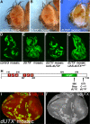
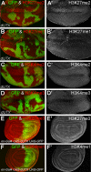
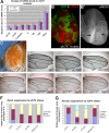
Trimethylated lysine 27 of histone H3 (H3K27me3) is an epigenetic mark for gene silencing and can be demethylated by the JmjC domain of UTX. Excessive H3K27me3 levels can cause tumorigenesis, but little is known about the mechanisms leading to those cancers. Mutants of the Drosophila H3K27me3 demethylase dUTX display some characteristics of Trithorax group mutants and have increased H3K27me3 levels in vivo. Surprisingly, dUTX mutations also affect H3K4me1 levels in a JmjC-independent manner. We show that a disruption of the JmjC domain of dUTX results in a growth advantage for mutant cells over adjacent wild-type tissue due to increased proliferation. The growth advantage of dUTX mutant tissue is caused, at least in part, by increased Notch activity, demonstrating that dUTX is a Notch antagonist. Furthermore, the inactivation of Retinoblastoma (Rbf in Drosophila) contributes to the growth advantage of dUTX mutant tissue. The excessive activation of Notch in dUTX mutant cells leads to tumor-like growth in an Rbf-dependent manner. In summary, these data suggest that dUTX is a suppressor of Notch- and Rbf-dependent tumors in Drosophila melanogaster and may provide a model for UTX-dependent tumorigenesis in humans.
Figures


Similar articles
-
An extracellular region of Serrate is essential for ligand-induced cis-inhibition of Notch signaling.Development. 2013 May;140(9):2039-49. doi: 10.1242/dev.087916. Development. 2013. PMID: 23571220 Free PMC article.
-
Drosophila Epsin's role in Notch ligand cells requires three Epsin protein functions: the lipid binding function of the ENTH domain, a single Ubiquitin interaction motif, and a subset of the C-terminal protein binding modules.Dev Biol. 2012 Mar 15;363(2):399-412. doi: 10.1016/j.ydbio.2012.01.004. Epub 2012 Jan 13. Dev Biol. 2012. PMID: 22265678 Free PMC article.
-
A Serrate-Notch-Canoe complex mediates essential interactions between glia and neuroepithelial cells during Drosophila optic lobe development.J Cell Sci. 2013 Nov 1;126(Pt 21):4873-84. doi: 10.1242/jcs.125617. Epub 2013 Aug 22. J Cell Sci. 2013. PMID: 23970418
-
Delivering the lateral inhibition punchline: it's all about the timing.Sci Signal. 2010 Oct 26;3(145):pe38. doi: 10.1126/scisignal.3145pe38. Sci Signal. 2010. PMID: 20978236 Review.
-
Cell and molecular biology of Notch.J Endocrinol. 2007 Sep;194(3):459-74. doi: 10.1677/JOE-07-0242. J Endocrinol. 2007. PMID: 17761886 Review.
Cited by
-
Histone demethylase UTX and chromatin remodeler BRM bind directly to CBP and modulate acetylation of histone H3 lysine 27.Mol Cell Biol. 2012 Jun;32(12):2323-34. doi: 10.1128/MCB.06392-11. Epub 2012 Apr 9. Mol Cell Biol. 2012. PMID: 22493065 Free PMC article.
-
Stress affects the epigenetic marks added by natural transposable element insertions in Drosophila melanogaster.Sci Rep. 2018 Aug 15;8(1):12197. doi: 10.1038/s41598-018-30491-w. Sci Rep. 2018. PMID: 30111890 Free PMC article.
-
Sudestada1, a Drosophila ribosomal prolyl-hydroxylase required for mRNA translation, cell homeostasis, and organ growth.Proc Natl Acad Sci U S A. 2014 Mar 18;111(11):4025-30. doi: 10.1073/pnas.1314485111. Epub 2014 Feb 18. Proc Natl Acad Sci U S A. 2014. PMID: 24550463 Free PMC article.
-
Drosophila UTX coordinates with p53 to regulate ku80 expression in response to DNA damage.PLoS One. 2013 Nov 12;8(11):e78652. doi: 10.1371/journal.pone.0078652. eCollection 2013. PLoS One. 2013. PMID: 24265704 Free PMC article.
-
Arabidopsis thaliana: a powerful model organism to explore histone modifications and their upstream regulations.Epigenetics. 2023 Dec;18(1):2211362. doi: 10.1080/15592294.2023.2211362. Epigenetics. 2023. PMID: 37196184 Free PMC article. Review.
References
-
- Agger, K., P. A. Cloos, J. Christensen, D. Pasini, S. Rose, J. Rappsilber, I. Issaeva, E. Canaani, A. E. Salcini, and K. Helin. 2007. UTX and JMJD3 are histone H3K27 demethylases involved in HOX gene regulation and development. Nature 449:731-734. - PubMed
-
- Bagchi, S., R. Weinmann, and P. Raychaudhuri. 1991. The retinoblastoma protein copurifies with E2F-I, an E1A-regulated inhibitor of the transcription factor E2F. Cell 65:1063-1072. - PubMed
-
- Baonza, A., and M. Freeman. 2005. Control of cell proliferation in the Drosophila eye by Notch signaling. Dev. Cell 8:529-539. - PubMed
-
- Blatch, G. L., and M. Lassle. 1999. The tetratricopeptide repeat: a structural motif mediating protein-protein interactions. Bioessays 21:932-939. - PubMed
-
- Boyer, L. A., K. Plath, J. Zeitlinger, T. Brambrink, L. A. Medeiros, T. I. Lee, S. S. Levine, M. Wernig, A. Tajonar, M. K. Ray, G. W. Bell, A. P. Otte, M. Vidal, D. K. Gifford, R. A. Young, and R. Jaenisch. 2006. Polycomb complexes repress developmental regulators in murine embryonic stem cells. Nature 441:349-353. - PubMed
Publication types
MeSH terms
Substances
Grants and funding
- GM074977/GM/NIGMS NIH HHS/United States
- R01 GM068016/GM/NIGMS NIH HHS/United States
- GM61672/GM/NIGMS NIH HHS/United States
- R01 GM081543/GM/NIGMS NIH HHS/United States
- R01 CA089455/CA/NCI NIH HHS/United States
- CA089455/CA/NCI NIH HHS/United States
- GM069905/GM/NIGMS NIH HHS/United States
- GM081543/GM/NIGMS NIH HHS/United States
- GM068016/GM/NIGMS NIH HHS/United States
- R01 CA150265/CA/NCI NIH HHS/United States
- R01 GM074977/GM/NIGMS NIH HHS/United States
- R01 GM069905/GM/NIGMS NIH HHS/United States
- R01 GM061672/GM/NIGMS NIH HHS/United States
LinkOut - more resources
Full Text Sources
Other Literature Sources
Molecular Biology Databases
