. 2010 Mar 23;107(12):5540-4.
doi: 10.1073/pnas.0912675107. Epub 2010 Mar 8.

Ablation of IL-17A abrogates progression of spontaneous intestinal tumorigenesis

Affiliations

Ablation of IL-17A abrogates progression of spontaneous intestinal tumorigenesis

Wook-Jin Chae et al. Proc Natl Acad Sci U S A. .

Abstract

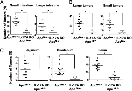
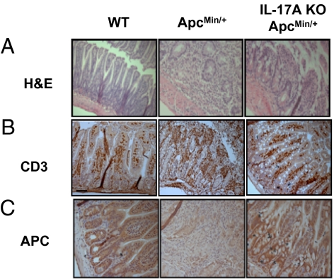
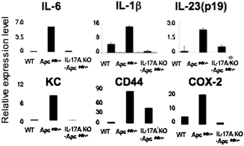
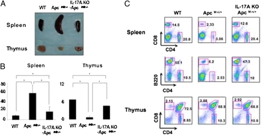
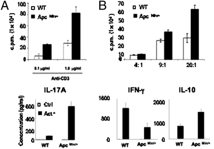
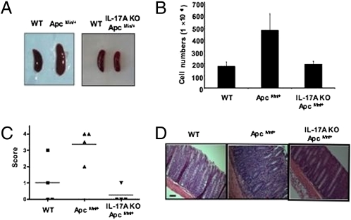
The intrinsic role of endogenous IL-17A in spontaneous intestinal tumorigenesis has not been addressed previously to our knowledge. Ablation of IL-17A significantly reduced tumor development in mice bearing a heterozygote mutation in the adenomatous polyposis coli (APC) gene (Apc(Min/+) mice). There was also a decrease in inflammatory cytokines and proinflammatory mediators, reduced infiltration of lymphocytes including T cells, and preservation of intestinal architecture and the presence of APC protein in intestinal epithelial cells. Interestingly, IL-17A ablation also corrected immunological abnormalities such as splenomegaly and thymic atrophy in Apc(Min/+) mice. CD4 T cells from Apc(Min/+) mice showed hyperproliferative potential in vitro and in vivo and increased levels of IL-17A and IL-10. The effector CD4 T cells from Apc(Min/+) mice were more resistant to regulatory T cell-mediated suppression. Finally, these CD4 T cells induced colitis in immunodeficient mice upon adoptive transfer, whereas the ablation of IL-17A in CD4 T cells in Apc(Min/+) mice completely abolished this pathogenic potential in vivo. Taken together, our results show that CD4 T cell-derived IL-17A promotes spontaneous intestinal tumorigenesis with altered functions of CD4 T cells in Apc(Min/+) mice.


Conflict of interest statement

The authors declare no conflict of interest.

Figures

Fig. 1.
Fig. 1.
IL-17A ablation significantly reduces intestinal tumorigenesis. (A) Tumor numbers of small and large intestine were counted in 20-week-old ApcMin/+ mice (n = 16) and IL-17A KO- ApcMin/+ mice (n = 13). (B) Tumors smaller than 3 mm and larger than 3 mm were counted separately. (C) Tumor numbers from each part of small intestine (duodenum, jejunum, and ileum). For both comparisons, *P < 0.002 using two-tailed t tests.
Fig. 2.
Fig. 2.
Ablation of IL-17A preserved small intestinal architecture in ApcMin/+ mice and reduces immune cell infiltration. Small intestines (ileum) of 20-week-old C57BL/6 (n = 5), ApcMin/+ (n = 4), and IL-17A KO- ApcMin/+ (n = 4) mice were harvested and stained with H&E (A) and murine anti-CD3 monoclonal antibody (B). Ileums of same group of mice were stained with anti-APC monoclonal antibody (C) with paraffin-embedded section.
Fig. 3.
Fig. 3.
IL-17A ablation reduced proinflammatory cytokines and mediators in tumors. Tumors (3 mm; one or two from each mouse) were dissected from 20-week-old ApcMin/+ mice (n = 5) and IL-17A KO- ApcMin/+ mice (n = 4). mRNA level was normalized against HPRT. For WT C57BL/6 mice, 5 mm of ileum was used. Values are means ± SD of all experiments.
Fig. 4.
Fig. 4.
Splenomegaly and thymic atrophy of ApcMin/+ mice are corrected by IL-17A ablation. Spleens and thymuses from 20-week-old ApcMin/+ and IL-17A KO ApcMin/+ mice and littermate controls of ApcMin/+ were dissected (A) and counted for cell numbers (B), and subsequently analyzed by flow cytometry (C). Flow cytometry is a representative of five mice per group. *P ≤ 0.002; one-way analysis was used with Tukey correction at an α of 0.05.
Fig. 5.
Fig. 5.
CD4 T cells from ApcMin/+ mice are functionally altered. (A) CD4 T cells from ApcMin/+ mice were isolated and activated with given amount of anti-CD3 and T cell–depleted irradiated splenocytes for 72 h. (B) Foxp3+ regulatory T cells were isolated from ApcMin/+ Foxp3-IRES-RFP mice and cocultured with Foxp3- CD4 effector T cells from either ApcMin/+ mice or their littermate control for 72 h for given ratios. (C) CD4 T cells from ApcMin/+ mice were isolated and activated for 96 h, and cytokines were measured by cytokine bead array. Data indicate means ± SD of three separate experiments as triplicates.
Fig. 6.
Fig. 6.
Ablation of IL-17A in CD4 T cells corrects altered functions in CD4 T cells from ApcMin/+ mice. (A) CD4 T cells from 11-week-old ApcMin/+ and IL-17A KO ApcMin/+ mice and littermate controls ApcMin/+ mice were isolated and transferred into Rag 2 KO mice. After 4 weeks of transfer, spleens were harvested and cell numbers were counted, and the ratio between CD4 effector T cells and Foxp3+ CD4 T cells was measured by flow cytometry (B). Colons were harvested and scored for their chronicity (C) and stained with H&E (D). The experiments are the representative of two independent experiments.

References

    1. Dubin PJ, Kolls JK. Th17 cytokines and mucosal immunity. Immunol Rev. 2008;226:160–171. - PubMed
    1. Ogura H, et al. Interleukin-17 promotes autoimmunity by triggering a positive-feedback loop via interleukin-6 induction. Immunity. 2008;29:628–636. - PubMed
    1. Abraham C, Cho J. Interleukin-23/Th17 pathways and inflammatory bowel disease. Inflamm Bowel Dis. 2009;15:1090–1100. - PubMed
    1. Atarashi K, et al. ATP drives lamina propria T(H)17 cell differentiation. Nature. 2008;455:808–812. - PubMed
    1. Korn T, Bettelli E, Oukka M, Kuchroo VK. IL-17 and Th17 Cells. Annu Rev Immunol. 2009;27:485–517. - PubMed

Publication types

MeSH terms

LinkOut - more resources