. 2010 Mar 23;107(12):5471-6.
doi: 10.1073/pnas.0908899107. Epub 2010 Mar 8.

Direct positive regulation of PTEN by the p85 subunit of phosphatidylinositol 3-kinase

Affiliations

Direct positive regulation of PTEN by the p85 subunit of phosphatidylinositol 3-kinase

Ryaz B Chagpar et al. Proc Natl Acad Sci U S A. .

Abstract

The phosphatidylinositol 3-kinase (PI3K) signaling pathway is deregulated in many human diseases including cancer, diabetes, obesity, and autoimmunity. PI3K consists of a p110 catalytic protein and a p85alpha regulatory protein, required for the stabilization and localization of p110-PI3K activity. The p110-PI3K enzyme generates the key signaling lipid phosphatidylinositol 3,4,5-trisphosphate, which is dephosphorylated by the PI3-phosphatase PTEN. Here we show another function for the p85alpha regulatory protein: it binds directly to and enhances PTEN lipid phosphatase activity. We demonstrate that ectopically expressed FLAG-tagged p85 coimmunoprecipitates endogenous PTEN in an epidermal growth factor dependent manner. We also show epidermal growth factor dependent coimmunoprecipitation of endogenous p85 and PTEN proteins in HeLa cells. Thus p85 regulates both p110-PI3K and PTEN-phosphatase enzymes through direct interaction. This finding underscores the need for caution in analyzing PI3K activity because anti-p85 immunoprecipitations may contain both p85:p110-PI3K and p85:PTEN-phosphatase enzymes and thus measure net PI3K activity. We identify the N-terminal SH3-BH region of p85alpha, absent in the smaller p55alpha and p50alpha isoforms, as the region that mediates PTEN binding and regulation. Cellular expression of p85DeltaSH3-BH results in substantially increased magnitude and duration of pAkt levels in response to growth factor stimulation. The ability of p85 to bind and directly regulate both p110-PI3K and PTEN-PI3-phosphatase allows us to explain the paradoxical insulin signaling phenotypes observed in mice with reduced PI3K or PTEN proteins. This discovery will impact ongoing studies using therapeutics targeting the PI3K/PTEN/Akt pathway.


Conflict of interest statement

The authors declare no conflict of interest.

Figures

Fig. 1.
Fig. 1.
p85 protein associates with PTEN in a growth factor-dependent manner in COS-1 cells, binds directly to PTEN and stimulates PTEN activity in vitro. (A) Domain structure of PTEN protein. (B) Coomassie blue stained SDS/PAGE of GST and GST-PTEN-C124S proteins immobilized on glutathione Sepharose beads and purified p85 protein used in pull-down experiments. (C) GST and GST-PTEN (C124S) were immobilized on glutathione Sepharose beads and mixed with purified p85 protein in a pull-down experiment. Bound p85 protein was detected by immunoblotting with anti-p85 antibody. As controls, pure p85 protein was loaded (0.05 μg) and GST-PTEN-C124S was incubated in the absence of p85. (D) Lysates from COS-1 cells transfected with FLAG-p85 ± EGF stimulation were immunoprecipitated (IP) and immunoblotted with the indicated antibodies. (E) Coomassie blue stained SDS/PAGE of purified His6-PTEN wild-type (WT) and phosphatase dead (C124S) proteins used for PTEN assays. (F) PTEN lipid phosphatase activity was assayed ± added p85, using a fluorescent lipid substrate and by resolving the reaction products by TLC. The His6-PTEN-C124S mutant lacks phosphatase activity. (G) Phosphatase activity was assayed over a range of PTEN (WT) concentrations. Results shown are the mean of three independent experiments ± SD (H) PTEN (0.56 μM) lipid phosphatase activity was determined with increasing concentrations of p85. (Inset) Lipid substrate alone (lane 1), lipid with p85 (1.12 μM; lane 2), and lipid with a high concentration of PTEN (3.2 μM; lane 3). Similar results were obtained in replicate experiments.
Fig. 2.
Fig. 2.
The BH domain of p85 binds PTEN. (A) Domain structure of p85 protein. (B) Schematic representation of the domains present in the FLAG-tagged p85 fusion proteins. (C and D) Lysates from COS-1 cells transfected with different FLAG-tagged domains of p85 were used in pull-down experiments with immobilized GST and GST-PTEN (C124S) as indicated.
Fig. 3.
Fig. 3.
The p85 protein binds PTEN via its N-terminal SH3-BH domains and p110 via its p110-binding (110B) domain. (A) Domains present in p85ΔSH3-BH protein. (B) Pull-down experiment with GST-PTEN-C124S and COS-1 lysates containing FLAG-p85ΔSH3-BH, a mutant lacking the N-terminal half of p85, is unable to bind PTEN. (C) Lysates (20 μg) from NIH 3T3 cells stably expressing near endogenous levels of different FLAG-p85 proteins ± PDGF were probed for pAkt (pS473) and total Akt. FLAG-p85 mutant expression levels were similar as determined using FLAG immunoblots. (D) Diagram illustrating regions of p85 involved in binding PTEN, p110, and activated RTKs.
Fig. 4.
Fig. 4.
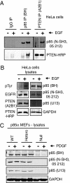
Growth factor dependent coimmunoprecipitation of endogenous p85 and PTEN from cells. (A) HeLa cells were serum-starved in 0.5% serum overnight and treated (+) or not (−) with EGF for 5 min. Cell lysates were immunoprecipitated and immunoblotted with the indicated antibodies. (B) Control HeLa cell lysates (20 μL) were immunoblotted with the indicated antibodies. (C) MEFs derived from p85α wild-type, heterozygous (+/−) and null (−/−) mice were serum-starved in 0.5% serum overnight and treated (+) or not (−) with PDGF for 5 min. Cell lysates (20 μL) were immunoblotted with the indicated antibodies. The p85 antibodies used were specific for p85α.
Fig. 5.
Fig. 5.
Resolving the paradox that both Pik3r1(+/−) mice and p85α(−/−) mice have insulin signaling phenotypes resembling PTEN(+/−) mice rather than p110α(+/−) mice. (A) PI3K signaling is transient in normal mice as a result of a balance between p85:p110-mediated generation of PI3,4,5P3 and p85:PTEN-mediated dephosphorylation of PI3,4,5P3. (B) p110α(+/−) mice contain half the normal amount of p110α. (C) PTEN(+/−) mice contain half the normal amount of PTEN. (D) Pik3r1(+/−) mice contain half the levels of p85α and p50α, both products of the Pik3r1 gene and both able to regulate p110-PI3K (and not p55α) as well as bind upstream activators. Decreased p85α levels do not, however, reduce p85:p110 and p85:PTEN complexes equally, with near normal levels of p85:p110 (19). The selective reduction of the p85 not associated with p110 includes p85:PTEN, specifically impairing PTEN-mediated PI3,4,5P3 dephosphorylation and leading to increased and sustained PI3K signaling. (E) Selective isoform-specific p85α(−/−) knockout mice do not express p85α but have normal levels of p50α, containing both the p110-binding domain and the pTyr-binding SH2 domains. Thus, p50α substitutes for p85α in its ability to link p110-PI3K to upstream pTyr activation signals. However, p50α lacks the N-terminal half of p85 (i.e., SH-BH domains), we have shown to be required for binding and positive regulation of PTEN activity. Thus, p50α is unable to bind and positively regulate PTEN resulting in increased and sustained PI3K signaling.

References

    1. Yuan TL, Cantley LC. PI3K pathway alterations in cancer: Variations on a theme. Oncogene. 2008;27:5497–5510. - PMC - PubMed
    1. Carracedo A, Pandolfi PP. The PTEN-PI3K pathway: Of feedbacks and cross-talks. Oncogene. 2008;27:5527–5541. - PubMed
    1. Samuels Y, Ericson K. Oncogenic PI3K and its role in cancer. Curr Opin Oncol. 2006;18:77–82. - PubMed
    1. Brachmann SM, Ueki K, Engelman JA, Kahn RC, Cantley LC. Phosphoinositide 3-kinase catalytic subunit deletion and regulatory subunit deletion have opposite effects on insulin sensitivity in mice. Mol Cell Biol. 2005;25:1596–1607. - PMC - PubMed
    1. Mauvais-Jarvis F, et al. Reduced expression of the murine p85alpha subunit of phosphoinositide 3-kinase improves insulin signaling and ameliorates diabetes. J Clin Invest. 2002;109:141–149. - PMC - PubMed

Publication types

MeSH terms

LinkOut - more resources