. 2010 Mar 23;107(12):5646-51.
doi: 10.1073/pnas.1001360107. Epub 2010 Mar 8.

Cardiac hypertrophy is not amplified by deletion of cGMP-dependent protein kinase I in cardiomyocytes

Affiliations

Cardiac hypertrophy is not amplified by deletion of cGMP-dependent protein kinase I in cardiomyocytes

Robert Lukowski et al. Proc Natl Acad Sci U S A. .

Abstract

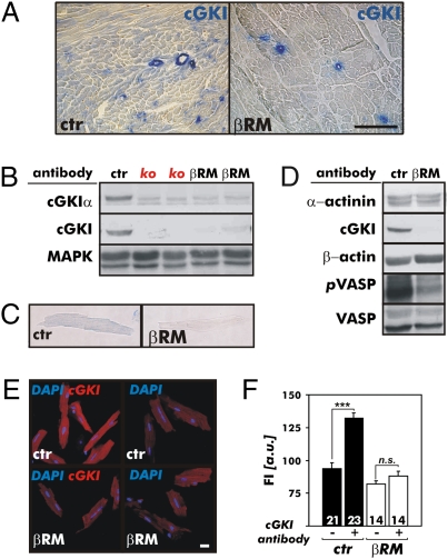
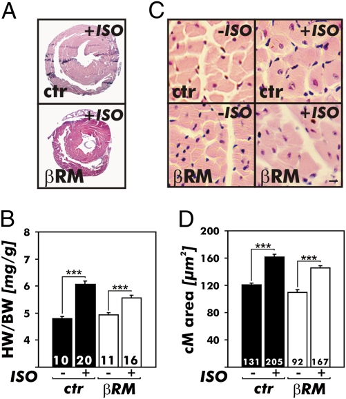
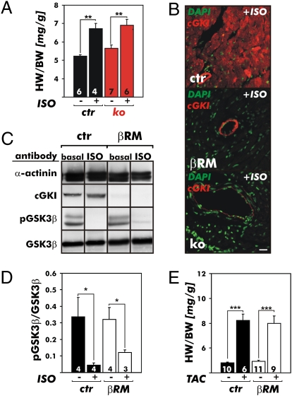
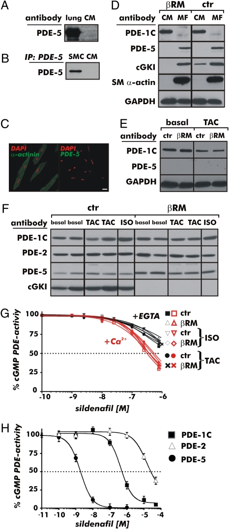
It has been suggested that cGMP kinase I (cGKI) dampens cardiac hypertrophy. We have compared the effect of isoproterenol (ISO) and transverse aortic constriction (TAC) on hypertrophy in WT [control (CTR)] mice, total cGKI-KO mice, and cGKIbeta rescue mice (betaRM) lacking cGKI specifically in cardiomyocytes (CMs). Infusion of ISO did not change the expression of cGKI in the hearts of CTR mice or betaRM but raised the heart weight by approximately 20% in both. An identical hypertrophic growth response was measured in CMs from CTR mice and betaRM and in isolated adult CMs cultured with or without 1 muM ISO. In both genotypes, ISO infusion induced similar changes in the expression of hypertrophy-associated cardiac genes and significant elevation of serum atrial natriuretic peptide and total cardiac cGMP. No differences in cardiac hypertrophy were obtained by 7-day ISO infusion in 4- to 6-week-old conventional cGKI-KO and CTR mice. Furthermore, TAC-induced hypertrophy of CTR mice and betaRM was not different and did not result in changes of the cGMP-hydrolyzing phosphodiesterase activities in hypertropic hearts or CMs. These results strongly suggest that cardiac myocyte cGKI does not affect the development of heart hypertrophy induced by pressure overload or chronic ISO infusion.


Conflict of interest statement

The authors declare no conflict of interest.

Figures

Fig. 1.
Fig. 1.
Expression analysis and activity of the cGKI in hearts of CTR mice and βRM. (A) By immunohistochemistry, cGKI was detected in the vasculature and myocardium of cardiac sections from adult CTR (ctr) mice but was absent from the heart muscle of βRM. In these animals, cGKI protein expression was limited to the SM layer of coronary vessels as expected by the endogenous SM22α gene promoter-driven expression of the cGKI construct. (Scale bar: 100 μm.) (B) Western blot analysis of ventricular protein lysates using polyclonal antibodies that specifically detect either the cGKIα isoform or all cGKI isoforms. The cardiac cGKIα isoform was present in heart lysates of the ctr mice but was not detected in hearts of conventional cGKI-KO (ko) mice or βRM. By identifying MAPK, equal loading of the gel is demonstrated. (C) Heart homogenates of ctr mice and βRM were subjected to immunoblotting analysis to show the strongly reduced cGKI-dependent phosphorylation of cardiac target proteins. The antibodies used to detect cGKI substrate phosphorylation were specific for the phospho-Ser239 residue of the phospho-VASP239 (pVASP). α-Actinin and VASP common antibodies were used as loading controls. cGKI immunohistochemistry (D) and immunofluorescence (E) of adult cardiac myocytes. The cGKI protein was only detectable in control cells but was not present in CMs of βRM. (Scale bar: 20 μm.) (F) Quantification of cGKI fluorescence intensity (FI) of adult ctr mice (black bars) and βRM (white bars) CMs. FI was determined using the specific cGKI antibodies and fluorophore-coupled secondary antibodies to detect the primary antibody protein complexes (***P < 0.001). a.u., arbitrary units; n.s., not significant.
Fig. 2.
Fig. 2.
Heart growth induced by ISO infusion for 7 days via implanted miniosmotic pumps in CTR mice and βRM. (A) Representative H&E-stained cardiac cross-sections of ISO-treated animals. Note that the hearts of βRM appear smaller because the animals weigh less [26.4 vs. 23.1 g for CTR (ctr) mice (n = 20) and βRM (n = 16), respectively]. (B) Chronic ISO infusion caused hypertrophy of the heart as indicated by increases of the HW/BW ratios. The hypertrophy response was similar in each genotype (**P < 0.01; ***P < 0.001). (C and D) Cross-sectional areas of cardiac myocytes. (C) H&E staining of heart sections from ctr mice and βRM at baseline (Left, −ISO) and after chronic ISO infusion (Right, +ISO). (Scale bar: 10 μm.) (D) Summary of cell size quantified by morphometry. ISO treatment provoked a significant increase in the CM (cM) cross-sectional areas in hearts of ctr mice and βRM. The areas were determined in the histological sections shown in A and as described in Materials and Methods (***P < 0.001).
Fig. 3.
Fig. 3.
Cardiac hypertrophy and signaling in various gene-targeted cGKI mouse models. (A) Effects of 7 days of chronic ISO infusion via implanted miniosmotic pumps on the hearts of 4- to 6-week-old CTR (ctr) mice and littermate conventional cGKI-KO (ko) mice. Chronic ISO infusion caused hypertrophy of the heart as indicated by increases of the HW/BW ratios. The response to the hypertrophy stimulus of each genotype indicated was similar in comparison to the respective saline-infused groups (**P < 0.01). (B) cGKI expression analysis in the hearts of CTR (ctr) mice, βRM, and ko mice that were chronically treated with ISO for 7 days. DAPI (green) was used as a nuclear counterstain. The cGKI protein (red) was detected in the vasculature and myocardium of ctr mice but was absent from the hearts of ko mice. In the βRM, the cGKI expression pattern was not influenced by the hypertrophy stimulus that induced a fetal gene program (Fig. S4) but was similar to baseline conditions and yet limited to the vasculature. (Scale bar: 20 μm.) Western blot (C) and quantification (D) of GSK3β-to-phospho-GSK3β (pGSK3β) ratio of ventricular protein lysates from ctr mice and βRM at baseline and after 7 days of ISO. (E) Effect of pressure overload induced by TAC on cardiac hypertrophy. Twenty-one days of TAC caused a significant increase in the HW/BW ratios in both genotypes analyzed in A (*P < 0.01; ***P < 0.001).
Fig. 4.
Fig. 4.
Expression and activity of cGMP-hydrolyzing PDEs in the hearts and isolated cardiac cells of CTR mice and βRM. The specificity of the PDE-5 antibodies used was verified by detecting the PDE-5 protein in the lung (A) and SM cells (SMCs) (B). Using the same antibodies, no PDE-5 protein was detectable in CMs. (C) In adult CMs from CTR mice, which were identified by antibodies specific for the cardiac marker protein α-actinin, PDE-5 was not detectable. (Scale bar: 20 μm.) (D) Western blot analysis of adult CMs and cardiac myofibroblasts (MFs) from CTR (ctr) mice and βRM. PDE-5 was absent from CMs but was detectable in MFs. SM α-actin was used to discriminate between MFs and CMs. The Ca2+/calmodulin-dependent cGMP- and cAMP-hydrolyzing PDE-1C was present only in CMs. (E) Western blot analysis of adult CMs purified previous to (basal) and after 21 days of TAC. (F) Expression of PDE-1/2/5 and the cGKI protein in different models of cardiac hypertrophy. No significant changes were detectable in the heart after TAC and/or ISO treatment between ctr mice and βRM as compared with the respective basal expression. (G) SIL inhibition of cGMP-hydrolytic activity in heart lysates at basal level and after ISO treatment or TAC. The IC50 of SIL on the cGMP-hydrolyzing PDE activity was similar for both genotypes and did not change as a result of the hypertrophic stimuli. Importantly, the IC50 determined was about 450 nM, and was therefore in the range of PDE-1C inhibition. (H) cGMP-hydrolyzing activity of different recombinant PDEs in the presence of SIL (100 pM to 100 μM). The IC50s for the recombinant enzymes were 1.9 nM, 422 nM, and 15 μM for PDE-5, PDE-1C, and PDE-2, respectively.

Comment in

References

    1. Nishikimi T, Maeda N, Matsuoka H. The role of natriuretic peptides in cardioprotection. Cardiovasc Res. 2006;69:318–328. - PubMed
    1. Oliver PM, et al. Hypertension, cardiac hypertrophy, and sudden death in mice lacking natriuretic peptide receptor A. Proc Natl Acad Sci USA. 1997;94:14730–14735. - PMC - PubMed
    1. Lopez MJ, et al. Salt-resistant hypertension in mice lacking the guanylyl cyclase-A receptor for atrial natriuretic peptide. Nature. 1995;378:65–68. - PubMed
    1. Knowles JW, et al. Pressure-independent enhancement of cardiac hypertrophy in natriuretic peptide receptor A-deficient mice. J Clin Invest. 2001;107:975–984. - PMC - PubMed
    1. Kishimoto I, Rossi K, Garbers DL. A genetic model provides evidence that the receptor for atrial natriuretic peptide (guanylyl cyclase-A) inhibits cardiac ventricular myocyte hypertrophy. Proc Natl Acad Sci USA. 2001;98:2703–2706. - PMC - PubMed

Publication types

MeSH terms