. 2010 Mar 23;107(12):5534-9.
doi: 10.1073/pnas.1000032107. Epub 2010 Mar 8.

Constraints within major histocompatibility complex class I restricted peptides: presentation and consequences for T-cell recognition

Affiliations

Constraints within major histocompatibility complex class I restricted peptides: presentation and consequences for T-cell recognition

Alex Theodossis et al. Proc Natl Acad Sci U S A. .

Abstract

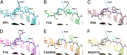
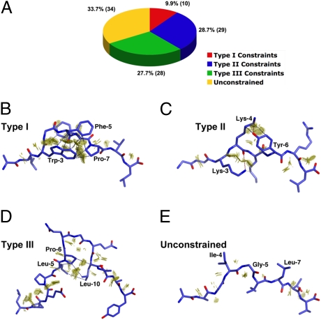
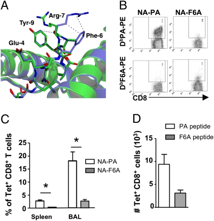
Residues within processed protein fragments bound to major histocompatibility complex class I (MHC-I) glycoproteins have been considered to function as a series of "independent pegs" that either anchor the peptide (p) to the MHC-I and/or interact with the spectrum of alphabeta-T-cell receptors (TCRs) specific for the pMHC-I epitope in question. Mining of the extensive pMHC-I structural database established that many self- and viral peptides show extensive and direct interresidue interactions, an unexpected finding that has led us to the idea of "constrained" peptides. Mutational analysis of two constrained peptides (the HLA B44 restricted self-peptide (B44DPalpha-EEFGRAFSF) and an H2-D(b) restricted influenza peptide (D(b)PA, SSLENFRAYV) demonstrated that the conformation of the prominently exposed arginine in both peptides was governed by interactions with MHC-I-orientated flanking residues from the peptide itself. Using reverse genetics in a murine influenza model, we revealed that mutation of an MHC-I-orientated residue (SSLENFRAYV --> SSLENARAYV) within the constrained PA peptide resulted in a diminished cytotoxic T lymphocyte (CTL) response and the recruitment of a limited pMHC-I specific TCR repertoire. Interactions between individual peptide positions can thus impose fine control on the conformation of pMHC-I epitopes, whereas the perturbation of such constraints can lead to a previously unappreciated mechanism of viral escape.


Conflict of interest statement

The authors declare no conflict of interest.

Figures

Fig. 1.
Fig. 1.
Peptide topology for the endogenous DPα and five APLs complexed with HLA-B*4402. In each panel, a cartoon representation is given of the Ag-binding cleft, whereas the bound peptide is presented in stick format. The α2-helix (residues 126–181) has been removed for clarity. In each of the APL panels, the index DPα peptide is superimposed in a semitransparent mode. Vdw interactions between atoms of P5 and other residues of the peptide are shown as blue dashes. (A) HLA-B*4402/DPα (1M6O; cyan carbon atoms). In addition to the interactions displayed, P5-Arg also interacts via its guanidinium head group with the side chain of Gln-155 (removed for clarity). (B) HLA-B*4402/DPα F3A (green carbon atoms). This APL displays an rmsd of 1.80 Å for all peptide atoms with respect to the index peptide and a maximum deviation of 3.27 Å in the side chain of P7-Phe. The side chain of P5-Arg is disordered beyond the Cγ atom. (C) HLA-B*4402/DPα R5A (red carbon atoms). Displayed is an rmsd of 0.24 Å for all peptide atoms with respect to the index epitope and a maximum deviation of 1.15 Å in the side chain of P8-Ser. (D) HLA-B*4402/DPα F7A (magenta). Rmsd for all peptide atoms is 1.06 Å. Maximum deviation is 5.61 Å in the side chain of P5-Arg. Interaction between P5-Arg and Gln-155 is maintained. (E) HLA-B*4402/DPα F3A/R5A (orange). All atom rmsd is 1.09 Å. Maximum deviation is 4.00 Å in the side chain of P5-Ala. (F) HLA-B*4402/DPα R5A/F7A (yellow). All atom rmsd is 0.47 Å. Maximum deviation is 1.59 Å in the side chain of P8-Ser.
Fig. 2.
Fig. 2.
MHC class-I restricted epitope self-constraints. (A) Summary of database mining results. Peptide ligands from 101 eligible pMHC-I entries in the PDB were assigned to one of three distinct constraints categories (i) type I, (ii) type II, (iii) type III, or alternatively were designated as being “unconstrained.” (BE) Examples of “self-constraints” displayed by MHC-I restricted peptides in the PDB database. Peptides are shown in stick format and the interacting vdw surfaces between residues are represented as a discontinuous surface of “contact dots” (dot density, 100 Å−2). Key interactions are further highlighted by blue (vdw) or red (H-bond) dashes. (B) “Type I” constraints observed in the 2.5-Å crystal structure of the p1049 9-mer peptide in complex with HLA-A*0201 (1B0G). (C) “Type II” constraints as observed in the 2.3-Å structure of the K7R mutant of the 8-mer HIV-1 GAG peptide in complex with HLA-B*0801 (1AGE). (D) “Type III” constraints displayed by the 1.6-Å structure of a 13-mer EBV antigen in complex with HLA-B*3501 (1ZHK). (E) The unconstrained 9-mer MART-1 peptide in complex with HLA-A*0201 at 1.9 Å (2GUO).
Fig. 3.
Fig. 3.
Loss of constrained interactions impacts on immunogenicity. (A) Comparison of the H2-Db/PA224 (blue) and H2-Db/PA224-F6A (green) crystal structures reveals that loss of constraints mediated by the aromatic ring of P6-Phe results in a shift of 8.5 Å in the position of the guanidinium head group of P7-Arg. Refined coordinates of the Ag-binding cleft are presented in cartoon format, whereas peptide atoms are shown as sticks. Intrapeptide vdw contacts involving P7-Arg are represented by blue dashes. Coordinates were superimposed using the Cα atoms of residues 1–180 of the heavy chain. (B) Lack of cross-reactivity between DbPA and DbPA-F6A epitopes. Naïve mice were infected with either NA-PA or NA-F6A PR8 influenza A viruses and lymphocytes from spleen and BAL were isolated 9 days later and costained with corresponding DbPA224 or DbPA-F6A –PE tetramer and anti-CD8a-FITC. Representative staining is shown. (C) Proportion of tetramer+CD8+ T cells from the spleen and BAL of mice infected with NA-PA (open bars) or NA-F6A (solid bars) PR8 viruses. Data are mean ± SD for groups of 6 mice. *, P < 0.001. (D) The F6A substitution within PA224 does not affect antigen presentation. Naïve mice were immunized s.c. with either PA224 (open bars) or PA-F6A (solid bars) peptide in CFA. Seven days later splenocytes were stained with either DbPA224 or DbPA-F6A –PE tetramer and anti-CD8a-PercPCy5.5. Shown is the absolute number of tetramer+CD8+ T cells (mean ± SD, n = 5 mice).

Similar articles

Cited by

References

    1. Bjorkman PJ, et al. Structure of the human class I histocompatibility antigen, HLA-A2. Nature. 1987;329:506–512. - PubMed
    1. Burrows SR, Rossjohn J, McCluskey J. Have we cut ourselves too short in mapping CTL epitopes? Trends Immunol. 2006;27:11–16. - PubMed
    1. Macdonald WA, et al. A naturally selected dimorphism within the HLA-B44 supertype alters class I structure, peptide repertoire, and T cell recognition. J Exp Med. 2003;198:679–691. - PMC - PubMed
    1. Rötzschke O, et al. Isolation and analysis of naturally processed viral peptides as recognized by cytotoxic T cells. Nature. 1990;348:252–254. - PubMed
    1. Burrows JM, et al. The impact of HLA-B micropolymorphism outside primary peptide anchor pockets on the CTL response to CMV. Eur J Immunol. 2007;37:946–953. - PubMed

Publication types

MeSH terms