

Visual field profile of optic neuritis: a final follow-up report from the optic neuritis treatment trial from baseline through 15 years
- PMID: 20212204
- PMCID: PMC4107874
- DOI: 10.1001/archophthalmol.2010.16
Visual field profile of optic neuritis: a final follow-up report from the optic neuritis treatment trial from baseline through 15 years
Abstract
Objective: To evaluate visual field abnormalities after an episode of optic neuritis among participants in the Optic Neuritis Treatment Trial.
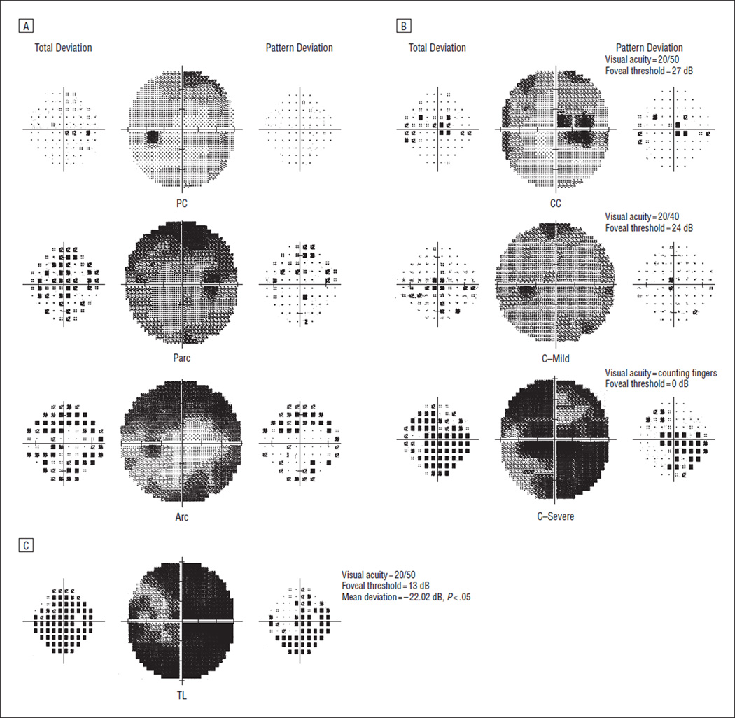
Methods: Three readers independently evaluated 10 443 visual fields from 454 patients and classified visual field abnormalities into 21 different monocular categories representing 3 general types of visual loss: diffuse, localized, and artifactual. Classification frequency was determined and reader agreement was evaluated. The association of visual field abnormality classifications with mean deviation, pattern standard deviation, visual acuity, and foveal threshold was assessed.
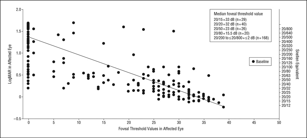
Results: At baseline, diffuse loss accounted for 66.2% of the abnormalities in the affected eyes but only 6.2% of the abnormalities in the fellow eyes. During years 1 through 15, the affected and fellow eyes exhibited predominantly localized loss in the nerve fiber bundle region (partial arcuate, paracentral, and arcuate defects). At year 1, 35.7% of the abnormalities in the affected eyes and 34.4% in the fellow eyes consisted of localized defects. At year 15, 39.5% of abnormalities in the affected eyes and 26.3% in the fellow eyes consisted of localized defects. Foveal threshold was highly correlated with visual acuity and contrast sensitivity in the affected eye at baseline (-0.82 vs 0.79, respectively), 6 months (-0.84 vs 0.81), and 1 year (-0.84 vs 0.79).
Conclusions: Diffuse and central loss were more predominant in the affected eye at baseline, and nerve fiber bundle defects (partial arcuate, paracentral, and arcuate) were the most predominant localized abnormalities in both the affected and fellow eyes during the study.
Figures
References
-
- Optic Neuritis Study Group. Visual function 15 years after optic neuritis: a final follow-up report from the Optic Neuritis Treatment Trial [published online November 5, 2007] Ophthalmology. 2008;115(6):1079.e5–1082.e5. - PubMed
-
- Volpe NJ. The Optic Neuritis Treatment Trial: a definitive answer and profound impact with unexpected results. Arch Ophthalmol. 2008;126(7):996–999. - PubMed
-
- Keltner JL, Johnson CA, Spurr JO, Beck RW Optic Neuritis Study Group. Baseline visual field profile of optic neuritis: the experience of the optic neuritis treatment trial. Arch Ophthalmol. 1993;111(2):231–234. - PubMed
